Download
1 / 12
130 likes | 148 Views
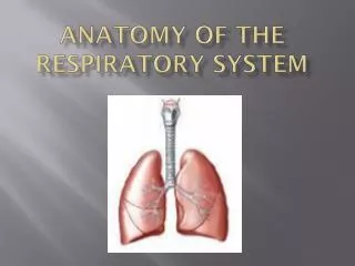
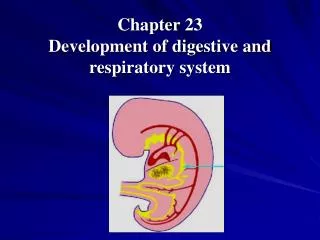
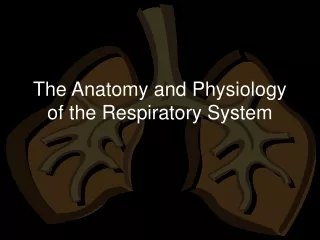
Chapter 23 Anatomy of the Respiratory System. Overview of the Respiratory System. The respiratory system functions as an air distributor and gas exchanger—supplying oxygen and removing carbon dioxide from cells (Figure 23-1)

E N D
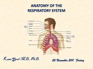
Overview of the Respiratory System • The respiratory system functions as an air distributor and gas exchanger—supplying oxygen and removing carbon dioxide from cells (Figure 23-1) • Alveoli—sacs that serve as gas exchangers; all other parts of respiratory system serve as air distributors • The respiratory system also warms, filters, and humidifies air • Respiratory organs influence speech, homeostasis of body pH, and olfaction • The respiratory system is divided into two divisions: • Upper respiratory tract—organs are located outside of the thorax and consist of nose, nasopharnyx, oropharnyx, laryngopharnyx, and larnyx • Lower respiratory tract—organs are located within the thorax and consist of trachea, bronchial tree, and lungs • Accessory structures include oral cavity, rib cage, and diaphragm
Upper Respiratory Tract • Nose • External portion of the nose consists of a bony and cartilaginous frame covered by skin containing sebaceous glands—the two nasal bones meet and are surrounded by the frontal bone to form the root; nose is surrounded by the maxilla (Figure 23-2) • Internal nose (nasal cavity) lies over the roof of the mouth, separated by the palatine bones • Cleft palate—palatine bones fail to unite completely and only partially separate the nose and the mouth, producing difficulty in swallowing • Cribriform plate—separates the roof of the nose from cranial cavity • Septum—separates nasal cavity into a right and a left cavity; consists of four structures: the perpendicular plate of the ethmoid bone, the vomer bone, the vomeronasal cartilages, and the septal nasal cartilage • Each nasal cavity is divided into three passageways: superior, middle, and inferior meati (Figure 23-3)
Upper Respiratory Tract • Nose (cont.) • Anterior nares—external openings to the nasal cavities, open into the vestibule • Sequence of air through nose into pharynx—anterior nares to vestibule to all three meati simultaneously to posterior nares • Nasal mucosa—a mucous membrane that air passes over; contains a rich blood supply (Figure 23-4) • Olfactory epithelium—specialized membrane containing many olfactory nerve cells and a rich lymphatic plexus • Paranasal sinuses—four pairs of air-containing spaces that open or drain into nasal cavity; each is lined with respiratory mucosa (Figure 23-5) • Nose is passageway for air traveling to and from lungs—filters the air, aids speech, and makes possible the sense of smell
Upper Respiratory Tract • Pharynx (throat) • Tubelike structure extending from the base of skull to the esophagus; made of muscle and divided into three parts (Figure 23-6): • Nasopharnyx • Oropharynx • Laryngopharnyx • Pharyngeal tonsils—located in the nasopharynx, called adenoids when they become enlarged • Oropharynx contains two pairs of organs—the palatine tonsils (most commonly removed) and the lingual tonsils (rarely removed) • Pharynx is pathway for respiratory and digestive tracts
Upper Respiratory Tract • Larynx (Figures 23-7 and 23-8) • Located between root of tongue and upper end of trachea • Consists of cartilages attached to each other by muscle and lined by a ciliated mucous membrane, which forms two pairs of folds (Figure 23-9): • Vestibular (false) vocal folds • True vocal cords • Framework of larynx is formed by nine cartilages: • Single laryngeal cartilages—the three largest cartilages: the thyroid cartilage, the epiglottis, and the cricoid cartilages • The paired laryngeal cartilages—three pairs of smaller cartilages: arytenoid, corniculate, and cuneiform cartilages
Upper Respiratory Tract • Larynx (cont.) • Muscles of larynx • Intrinsic muscles both insert and originate within larynx • Extrinsic muscles insert in larynx but originate on some other structure • Larynx functions as part of the airway to the lungs and produces the voice
Lower Respiratory Tract • Trachea (windpipe) extends from the larynx to the primary bronchi (Figure 23-11) • Furnishes part of the open airway to the lungs—obstruction causes death
Lower Respiratory Tract • Bronchi and alveoli • Lower end of trachea divides into two primary bronchi, one on right and one on left, which enter lung and divide into secondary bronchi that branch into bronchioles, which eventually divide into the alveolar ducts (Figure 23-13) • Alveoli are the primary gas exchange structures • Respiratory membrane—the barrier between which gases are exchanged by alveolar air and blood (Figure 23-16) • Respiratory membrane consists of alveolar epithelium, capillary endothelium, and their joined basement membranes • Surfactant—a component of the fluid coating the respiratory membrane that reduces surface tension • Bronchi and alveoli distribute air to lung’s interior
Lower Respiratory Tract • Lungs • Cone-shaped organs extending from the diaphragm to above the clavicles (Figure 23-18) • Hilum—slit on lung’s medial surface where primary bronchi and pulmonary blood vessels enter • Base—inferior surface of lung that rests on diaphragm • Costal surface—lies against ribs • Left lung is divided into two lobes—superior and inferior • Right lung is divided into three lobes—superior, middle, and inferior • Lobes are further divided into functional units—bronchopulmonary segments • Ten segments in right lung • Eight segments in left lung • Lungs have two functions—air distribution and gas exchange
Lower Respiratory Tract • Thorax (Figure 23-19) • Thoracic cavity has three divisions divided by the pleura: • Pleural divisions—part occupied by lungs • Mediastinum—part occupied by esophagus, trachea, large blood vessels, and heart • Thorax functions to bring about inspiration and expiration
Cycle of Life: Respiratory System • Respiration may be affected by developmental defects, age-related structural changes, or loss of function throughout the life cycle • Age-related changes affect vital capacity, make ventilation difficult, or reduce the oxygen– or carbon dioxide–carrying capacity of blood • Respiratory efficiency is reduced in old age as a result of changes in ribs, respiratory muscles, and hemoglobin levels