Download
1 / 55
570 likes | 736 Views
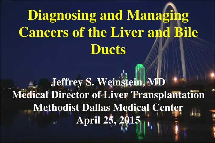
Diagnosing and Managing Cancers of the Liver and Bile Ducts Jeffrey S. Weinstein, MD Medical Director of Liver Transplantation Methodist Dallas Medical Center April 25, 2015. Types of Malignant Liver Tumors. Metastatic tumors (most common) Hepatocellular carcinoma (HCC)

E N D
Diagnosing and Managing Cancers of the Liver and Bile DuctsJeffrey S. Weinstein, MDMedical Director of Liver Transplantation Methodist Dallas Medical CenterApril 25, 2015
Types of Malignant Liver Tumors • Metastatic tumors (most common) • Hepatocellular carcinoma (HCC) • Cholangiocarcinoma (CCA) • Combined HCC/CCA • Fibrolamellar carcinoma • Hepatoblastoma • Cystadenocarcinoma • Epitheliod hemangioendothelioma • Angiosarcoma
Hepatocellular (HCC) • Clinical features • Diagnostic studies • Staging • Treatment options • Role of transplant
Clinical Features of HCC • Usually occur in background of chronic • liver disease and cirrhosis • Small tumors usually cause no symptoms • Larger tumors can be associated with: • - abdominal pain • - loss of appetite and weight loss • - worsening liver function • - fever • - jaundice • - intra-abdominal bleeding
Diagnostic studies for HCC • Labs • - AFP tumor marker • Radiologic studies • - Ultrasound • - Multiphase MRI • - Multiphase CT • Biopsy
AFP Limitations and Uses • Limitations • - may be elevated with chronic hepatitis • - elevated in HCC in only about ~ 40% • Uses • - level > 20 useful for screening but adds to costs • and false positive rates • - level > 400-500 about 99% specific • - level > 400-500 have worse prognosis
Non-Invasive Radiologic Studies Used for HCC • Ultrasound • - screening and staging • Multiphase contrast enhanced CT or MRI of • abdomen and pelvis • - screening, diagnosis, and staging • Chest CT +/- Bone scan • - staging
Invasive Studies Used for HCC • Ultrasound or CT guided biopsy • - diagnosis and staging • Endoscopic ultrasound • - diagnosis and staging • Angiography • - diagnosis and treatment
HCC findings on Contrast Enhanced MRI or CT • Requires dedicated 4-phase study • - Early and late arterial phases • - Portal venous phase • - Late venous phase • Typical HCC • - early arterial enhancement • - late venous washout
MRI Appearance of HCC Arterial phase Portal venous phase Late venous phase
Diagnostic Approach to HCC Mass < 1cm on Ultrasound Repeat Ultrasound every 3- 6 months for 2 years 0r MRI/CT x 1 No growth Growth in mass Resume surveillance every 6 months Typical vs. Atypical for HCC Contrast enhanced CT or MRI
Diagnostic Approach to HCC Mass < 1cm on MRI/CT Monitor with U/S q 3 months Typical for HCC Atypical for HCC Growth in mass Treat for HCC Repeat MRI/CT Atypical for HCC Check CA 19-9, biopsy or resect mass
Diagnostic Approach to HCC Mass > 1cm on MRI/CT Typical for HCC Atypical for HCC Obtain other study MRI/CT Treat for HCC Atypical for HCC Check CA 19-9, biopsy or resect mass
Tumor Staging for HCC • Critical for determining treatment options • Based on: • 1) Size and number of tumors • 2) Extension of tumor into adjacent structures • 3) Presence of metastases • 4)Severity of underlying liver disease • 5) Overall physical condition of the patient
Staging Severity of Liver Disease Child Pugh Score Ascites Bilirubin level Hepatic encephalopathy Albumin level Prothrombin time/INR MELD score Prothrombin time/INR Bilirubin level Serum creatinine Signs of Portal Hypertension Varices Low platelets Ascites Enlarged spleen
Treatment Options for HCC • Surgical resection • Liver transplant (OLTx) • Microwave or radiofrequency ablation • Transarterial chemoembolization (TACE) • Transarterial radioembolization • Radiation and stereotactic radiotherapy • Systemic chemotherapy • Cryoablation • Percutaneous alcohol ablation
Surgical Resection for HCC • Non-cirrhotic liver or well-compensated liver • disease • Lesion size and location amenable to surgery • Tumor confined to the liver • No invasion of large vessels
UNOS Guidelines for OLTx & HCC • Tumor < 5cm or three tumors all < 3cm • Surgical resection not possible • Tumor confined to liver • No invasion of large vessels or lymph nodes • Cirrhotic liver • No other contraindications to OLTx • Candidates allocated MELD score of 22 • MELD upgrade every 3 months if UNOS • criteria remain fulfilled
OLTx and HCC • Bridging therapies if waiting time expected to • be 6 months or more • - MWA or RFA for tumors < 3 cm • - TACE or Radioembolization for larger or • multiple tumors • About 20% will be removed from list for tumor • progression before OLTx • 5 year survival after OLTx ~ 75%
Locoregional Therapies for HCC • MWA or RFA for tumors < 3cm • TACE, Radioembolization and Stereotactic • radiotherapy for larger or multiple tumors • Tumor or tumors confined to liver • Absence of severe liver dysfunction • Reduce tumor burden before resection • Bridging therapy before OLTx • Primary treatment for those who are not • candidates for surgery or OLTx
Systemic Therapies for HCC • For advanced tumors and intact liver function • Vascular endothelial growth factor inhibitors • - Sorafenib* • - Bevacizumab • Epidermal growth factor inhibitors • - Erlotinib • - Cetuximab • Chemotherapy • - Gemcitabine + Cisplatin or Oxaliplatin • - Low dose Doxorubicin or Capecitabine
Cholangiocarcinoma (CCA) • Diagnostic studies • Staging • Treatment options • Unique issues related to PSC • Role of liver transplant (OLTx)
Anatomic Classification for CCA • Intrahepatic 10% • - arise from intrahepatic biliary tree • Distal 40% • - arise below the cystic duct insertion • Perihilar 50% (most common in PSC) • - Bismuth-Corlette classification • - Klatskin tumor any involvement of hepatic duct • bifurcation
CCA and PSC • Incidence of 0.6 to 1.5% per year • Lifetime risk 5 t0 15% • Younger age of onset ~ 30-50 than with CCA • not associated with PSC ~ 50-70 • Most tumors are perihilar • 1/3 of cases diagnosed within 2 years of • when PSC is first diagnosed • Smoking and alcohol use may increase risk
When to Suspect CCA in Patients with PSC • Rapid deterioration in clinical condition for • those with PSC • Jaundice, dark urine, pale stools, itching • Dull RUQ pain • Weight loss • Rising Alkaline phosphatase and bilirubin • Elevated CA 19-9 (> 100)
Diagnosing CCA • Clinical and laboratory abnormalities • Elevated tumor markers – CA 19-9 • Radiologic changes on MRCP, MRI/CT or • PET scan • Endoscopic findings on ERCP or EUS • Tumor cells identified on brushings or biopsy • Findings at time of Laparoscopy or Surgery
Studies Used to Diagnose CCA • Serum tumor markers • - CA 19-9 • Radiologic studies • - Ultrasound (U/S), MRCP, CT, MRI, and PET scans • Interventional studies • - ERCP and Endoscopic Ultrasound (EUS) • - Percutaneous cholangiogram (PTC) • - CT/MRI guided biopsies
CA 19-9 Limitations and Uses • Limitations • - cannot be detected in 5-10% of patients • - may be elevated in other conditions • pancreatic CA, acute cholangitis, or biliary obstruction • Uses • - level > 1000 correlated with advanced tumors • - level > 37 useful for screening but poor specificity • - level > 100-129 useful for screening or in patients • with suspicious stricture • - monitoring response to therapy
Non-Invasive Radiologic Studies Used for CCA • Ultrasound +/- doppler • screening and staging • MRCP • screening, diagnosis, and staging • Contrast enhanced multiphase CT or MRI • diagnosis and staging • PET/CT scan • diagnosis and staging
Radiologic Changes in PSC with Suspected CCA • Stricture progression • “Dominant stricture” • Increased dilation of the biliary tree proximal • to stricture • Polypoid mass within the bile duct
CCA Changes on MRCP Radiologic Changes in PSC with Suspected CCA
Radiologic Procedures and Interventions for CCA • Percutaneous cholangiography • - diagnose & stage tumor, alleviate obstruction • - assess, brush, biopsy, and stent strictures • - more difficult to do in PSC • CT/MRI guided biopsies • - confirm lymph node involvement with tumor • - confirm suspected primary tumor* • * risk of tumor seeding with biopsies
Endoscopic Procedures and Interventions for CCA • ERCP +/- cholangiography or intraductal U/S • - diagnose & stage tumor, alleviate obstruction • - assess extent of tumor in and around bile duct • - visualize, brush, and biopsy suspected tumors • - stent obstructing tumors • - has risk for infection (cholangitis) • Endoscopic ultrasound • - diagnose & stage tumor • - assess tumor extent, regional lymph nodes and vessels • - biopsy tumor* or regional lymph nodes • * risk of tumor seeding when tumor biopsied
CCA on Cholangiography Ulcerated stricture Ulcerated and nodular stricture
Diagnostic Challenges Related to CCA in Patients with PSC • Symptoms of PSC progression can mimic that • of tumor • CA 19-9 may be elevated from acute • cholangitis or benign causes for impaired • biliary excretion • Mass lesions less common • Biliary dilation proximal to point of • obstruction is less common
Tumor Staging for CCA • Critical for determining treatment options • Based on: • 1) Extent of tumor • 2) Severity of underlying liver disease • 3) Overall physical condition of the patient • Laparoscopy and/or open surgery often • required to complete the staging process
Treatment Options for Extrahepatic CCA • Surgery for patients without PSC • Transplant for select patients with PSC • Chemoradiotherapy • Systemic chemotherapy • Intra-arterial chemotherapy • Intra-arterial radiotherapy • Photodynamic therapy
CCA and PSCWhat’s the Difference? • Bile duct damage present in all patients • Underlying liver disease present in most patients • Diagnostic challenges • - benign vs. malignant bile duct stricture • - similar lab, radiologic, & endoscopic abnormalities • Surgical limitations • - recurrent infections • - cirrhosis and liver dysfunction • Cancer screening for CCA possible in PSC
Treatment of CCA in Patients With PSC • Stage and diagnose tumor with radiologic and • endoscopic studies • For large, locally advanced or metastatic tumors • - Nonsurgical treatments as for patients without PSC • - OLTx not recommended • For suspected tumor that is small and confined • - Needle biopsy should be avoided • - Refer to transplant center with protocol for CCA
Selecting Patients With PSC and CCA for OLTx • Must be an acceptable candidate for transplant • No intrahepatic or extrahepatic metastases • Diagnosis of CCA based on: • - Positive bushing or transcatheter biopsy or • - CA 19-9 > 100 with a suspicious mass or stricture • on imaging studies or cholangiography or • - Abnormal cells by FISH with suspicious stricture • Pre-OLTx treatment with chemoradiation • Staging laparotomy to exclude metastatic disease
OLTx for CCA in Patients With PSC • Complete treatment with chemoradiation and • have no metastases on staging laparotomy • MELD score 22 -> increased every 3 months if • criteria for transplant still met • Acceptable outcomes • Mayo Clinic experience • - 82%, 63% and 55% survival at 1, 3, and 5 years • Multicenter experience • - 68% and 53% survival at 2 and 5 years
Treatment of Locally Advanced CCA not Amenable to OLTx • Chemoradiotherapy • Photodynamic therapy • Systemic chemotherapy • Percutaneous or endoscopic biliary stents for • those with obstructive jaundice • Prophylactic antibiotics in those with history • of bacterial cholangitis
Radiation Therapies for CCA • External Beam Radiation (EBRT) * • High dose radiation therapies * • - Transcatheter brachytherapy +/- EBRT • - 3-Dimmensional conformal RT • - Intensity modulated RT • Stereotactic radiotherapy • * Radiation treatments typically combined with • chemotherapy with 5-FU or Capecitabine
Treatment of Advanced CCA • Good performance status • - Gemcitabine + Cisplatin or Oxaliplatin (1st line) • - Gemcitabine + Capecitabine • - Capecitabine + Oxaliplatin • - Ertonilab + Bevacizumab • Borderline performance status • - Gemcitabine or Capecitabine or 5-FU +Leucovorin • Poor performance status • - no chemotherapy • Stents for obstruction and antibiotics for cholangitis
Management of CCA in PSC CCA Diagnosed Advanced tumor Localized tumor Refer for OLTx Chemoradiotherapy Staging Laparotomy Chemoradiotherapy Photodynamic therapy and/or Chemotherapy Metastatic tumor Yes No List for OLTx
Specific Cancer Risks in PSC • Colon cancer • Gallbladder cancer • Hepatocellular carcinoma (HCC) in • those with underlying cirrhosis • Cholangiocarcinoma (CCA)