Download

1 / 44
550 likes | 957 Views
Malignant bone tumors. Primary Secondary (metastasis). Primary malignant tumors. Multiple myeloma Osteosarcoma Ewing sarcoma Chondrosarcoma. Uncommon cancer Most commonly affects the long bones 20 % of pediatric bone tumors are malignant.

E N D
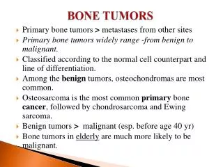
Malignant bone tumors Primary Secondary (metastasis)
Primary malignant tumors Multiple myeloma Osteosarcoma Ewing sarcoma Chondrosarcoma
Uncommon cancer • Most commonly affects the long bones • 20 % of pediatric bone tumors are malignant. • 66% of adult bone tumors are malignant, most commonly mets. • The most common type of bone cancer in adults is metastatic cancer from other organs
Primary bone cancer Risk factors: • Radiotherapy & chemotherapy • Paget's disease • Family Hx(hereditary retinoblastoma) Signs & symptoms • Fever, Night sweats, Fatigue & Unintended weight loss • Bone pain that often is nocturnal • Swelling & tenderness near the affected area • Pathological fractures
Osteosarcoma • Most common primary bone malignancy • Incidence: 2.8 per million • M >F • Age 10-25 years (the 8th most common childhood cancer) ** Prognosis • Aggressive tumor • Metastasis to the lung • 5-year survival • Without mets is 70% • With mets is 25%
Where Mainly affects metaphysis of long bones More in: • Knee • Distal femur • Upper tibia • Humerus (prox.end) • Maxilla
Clinical features • Pain: • Dull aching • Progressive • Constant • Worse at night • Swelling • Redness • Hotness • Tenderness • Pathological fracture
DDX • Stressfracture • Ewing's sarcoma • Osteomyelitis • Osteochondroma • Osteoblastoma • Bone cysts • Chondroblastoma • Chondrosarcoma • Giant cell tumor
Diagnosis: ** history & physical examination • Radiological studies: • X - ray • CT • Bone scan & MRI • Bone biopsy, the only definitive method to determine whether a tumor is malignant or benign. Treatment: • Surgical resection • Preoperative & postoperative chemotherapy
X-ray findings • Lesion • Cortical destruction • Extension to the marrow or soft tissue • Codman’s triangle a term used to describe the triangular area of new subperiosteal bone that is created when a lesion, often a tumour, raises the periosteum away from the bone. • Sunburst Effect Osteosarcomas can be • Predominantly osteolytic • Predominantly osteoblastic • Mixture
Clinical appearance of a teenager who presented with osteosarcoma of the proximal humerus • Swelling throughout the deltoid region • Disuse atrophy of the pectoral muscule
White arrow: codman triangle • Black arrow: soft tissue mass
Paget’s sarcoma • Paget’s disease of bone occasionally undergoes malignant transformation; most osteosarcomas appearing after the age of 50 years fall into this category. • This tumour is more malignant than classic osteosarcoma • Most patients have pulmonary metastases by the time the tumour is diagnosed. • Even with radical resection or amputation and chemotherapy the 5-year survival rate is low. If the lesion is definitely extra compartmental, palliative treatment by radiotherapy may be preferable; chemotherapy is usually difficult because of the patient’s age and uncertainty about renal and cardiac function.
Ewing sarcoma • A malignant round-cell tumor. • Rare disease (incidence 0.6 per million • 2nd most common bone malignancy in pediatrics. • M>F • Age 10-20 years • Usually the lesions are diaphyseal • Mets (30%), most commonly in the lungs & other bones & less commonly in the bone marrow.
Most common areas: • Pelvis • Femur • Humerus • Ribs • Clavicle
Clinical feature: • Pyrexia • Pain: • Constant • Increase with movement • Limping • Swelling, warm, tender & red Radiological studies: • X-Ray • Lyticmedullary lesion • Onion skin appearance • CT-scan • Bone scan & MRI
White arrow: onion skin apperance • Red circle: sunburstperiosteal reaction • Blue circle: osteolytic lesion
Periosteal reaction • Osteolytic lesion
Treatment: • Local radiotherapy combined with systemic chemotherapy • In young children amputation may be necessary due to severe compromise of bone growth. • Prognosis, 5-year survival • 50% with the 1st approach • 75% with the 2nd approach
Multiple myeloma ○ Malignant tumor of plasma cells. → originate from bone marrow ○ Most common non-metastatic malignant bone tumor ○ Patients present to orthopedics clinic with back, shoulder or hip pain. and pathological fracture , Spine is the most common location for a pathological fracture.
■ Hypercalcaemia may cause symptoms such as thirst, polyuria and abdominal pain. ■ Associated features of the marrow cell disorder are plasma protein abnormalities, increased blood viscosity and anaemia. Bone resorption leads to hypercalcaemia in about one-third of cases. ■ Late secondary features are due to renal dysfunction and spinal cord or root compression caused by vertebral collapse.
○ Increase in: AP “alkaline phosphatase”/ESR/calcium (leading to renal stones). ○ Anemia: antibodies against RBC’s. ○ Amyloidosis: in heart and kidney. ○ On electrophoresis: M-bands spike (50% IgG, 25% IgA) ○ Bence Jones proteins in urine.
X-rays often show nothing more than generalized osteoporosis; but remember that myeloma is one of the commonest causes of osteoporosis and vertebral compression fracture in men over the age of 45 years. • Moth eaten appearance on X-ray. • Bone scan: sometimes gives negative results (30%) → so we should whole skeletal survey
Management: chemo and radiotherapy (highly responsive), in addition to Bisphosphonates to decrease calcium. • median survival rate of only 2–5 years.
Most common malignant lesion of the bone. • The commonest source is carcinoma of the breast; next in frequency are carcinomas of the prostate, kidney, lung, thyroid, bladder and gastrointestinal tract. • Carcinomas are much more likely to metastasize to bone than sarcomas • Typically multifocal BUT renal and thyroid carcinomas produce only a solitary lesion. • Common sites for metastasis are vertebrae, pelvis, proximal parts of the femur & humerus. • Mets: • Direct extension • Retrograde venous flow • Seeding with tumor emboli via the blood circulation
Mets (adults) • Osteoblastic behaviour • Prostate • Stomach • Bladder • Breast • Osteolyticbehaviour • Lung • Kidney • Colon • Thyroid • Breast
Destructive expanded osteolytic lesion in the metacarpal of the thumb in a 55-year-old man with lung carcinoma.
Typical x-ray appearance of osteolytic bone metastases. This plain pelvic x-ray film of a 75-year-old patient with breast carcinoma shows multipleosteolytic bone lesions. =>decrease in bone density.
Typical x-ray appearance of osteoblastic bone metastases. This plain pelvic x-ray film of a patient with prostate cancer shows multipleosteoblastic metastases to the pelvis and lumbar (L4) and sacral (S1) vertebral bodies.=>increase in bone density
Mets (kids) • Neuroblastoma • Wilm’s tumor • Osteosarcoma • Ewing’s sarcoma • Rhabdomyosarcoma
Presentation • Pain is the commonest which results in reduced mobility, and often the only clinical feature • The sudden appearance of backache or thigh pain in an elderly person (especially someone known to have been treated for carcinoma in the past) is always suspicious. • Bone weakness which predispose to pathologic fractures. • Palpable masses (large bony lesions). • Neurologic impairment due to spinal epidural compression. • Anemia (decreased red blood cell production) is a common blood abnormality in these patients.
Approach • History & physical examination • Radiological studies • Plain X-ray • MRI • CT scan • Bone scan (Technetium-99m) • Laboratory studies • Biopsy
Radiological studies • X-ray: destruction of bone and/or lucent Lesions of Bone • Bone scan: most cost-effective and available whole-body screening test for the assessment of bone metastases. • CT • Useful in evaluating suspicious bone scintiscan findings • Useful in guiding needle biopsy, particularly in vertebral lesions. • MRI • Useful in evaluating suspicious bone scintiscan findings • Help in detecting metastatic lesions before changes in bone metabolism • Helpful in determining the extent of local disease in planning surgery or radiation therapy.
Treatment ■ treatment is entirely symptomatic. ■ Most patients require analgesics. ■ radiotherapy is used both to control pain and to reduce metastatic growth. ■ Treatment of fractures. ■ Prophylactic fixation. ● Large deposits that threaten to result in fracture should be treated by internal fixation while the bone is still intact. ■ Spinal stabilization ● Vertebral fractures usually require some form of support.
Treatment • Radiation therapy combined with chemotherapeutic or hormonal agents, is the most common treatment modality. • Early use of radiation and bisphosphonates (zoledronic acid, pamidronate) slows bone destruction. • Some tumors are more likely to heal after radiation therapy: • Blastic lesions of prostate and breast • Lytic destructive lesions of lung and renal cell • Surgery is indicated in fractures or large metastatic mass.