Download
1 / 21
430 likes | 2.82k Views
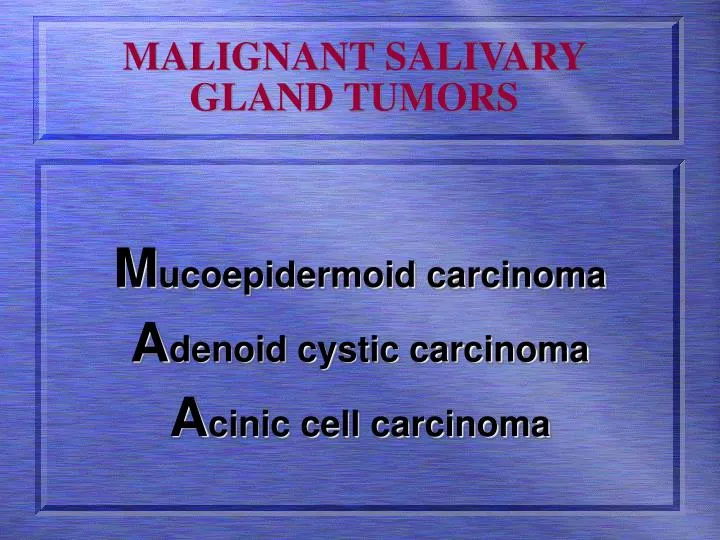
MALIGNANT SALIVARY GLAND TUMORS . M ucoepidermoid carcinoma A denoid cystic carcinoma A cinic cell carcinoma. Mucoepidermoid Carcinoma.

E N D
MALIGNANT SALIVARY GLAND TUMORS Mucoepidermoid carcinoma Adenoid cystic carcinoma Acinic cell carcinoma
Mucoepidermoid Carcinoma • Malignant salivary gland tumor is of varying degree of aggressiveness composed of mucous secreting and stratified squamous epithelial cells and lacking a capsule.
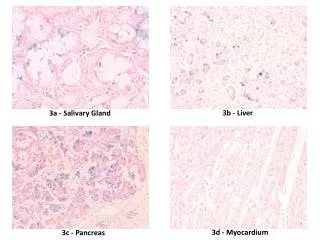
Mucoepidermoid Carcinoma CLINICAL FEATURES: • Adulthood tumor • Significant female predilection SITE: • 50% MEC occur in the parotid gland (arising in superficial lobe) • 20% occur on the palate • Rest of the lesions arising from the minor salivary glands with the buccal mucosa, lips, tongue and retro molar areas to be the favored sites. C/F.. Cont..d
Mucoepidermoid Carcinoma • The tumor may be movable, which is an uncommon feature for a malignant tumor. High grade lesions are often fixed to the adjacent tissues. • Their size is 1-4 cm when diagnosed. • There may be facial weakness due to VII nerve involvement.
Mucoepidermoid Carcinoma • HISTOPATHOLOGY: • They have three dominant cell types • Mucinous, epidermoid and intermediate • Their cells are arranged in the nests and diffuse sheets that may surround cystic spaces • There is no capsule, but the edge of the tumor is well-demarcated
Mucoepidermoid Carcinoma • There may be focal areas of malignant cells infiltrated into the normal salivary tissue • Tumors predominant mucous cells and more cystic spaces are classified as low grade (with limited metastatic potential) • Those with solid sheets and fewer mucous secreting cells and high proportion of stratified squamous epithelium are classified as high grade tumors
Mucoepidermoid CarcinomaLOW GRADE / HIGH GRADE
Mucoepidermoid Carcinoma TREATMENT/PROGNOSIS: • Low grade tumors follow a benign course whereas high grade show distant metastasis to the regional lymph nodes as well ( cervical lymph nodes) • Treatment of primary malignancy is managed with surgery followed by radiotherapy to the primary site. • Prognosis depends on the histological grade of the malignancy.
ADENOID CYSTIC CARCINOMA • It is a malignant salivary gland tumor composed of cuboidal cells in a solid cribriform tubular pattern • ACC is one of the most deceptive and frustrating tumor of the head and neck region ORIGION: • ACC arises from intercalated duct reserve cells or the terminal tube complex
ADENOID CYSTIC CARCINOMA CLINICAL FEATURES: • Peak incidence is in sixth decade of life with slight female predilection • 50-70% cases reported are in the minor salivary glands, the major glands that are affected are the parotid glands • In major salivary glands, the clinical appearance is that of a unilocular mass, which is firm on palpation • There might be some pain and tenderness
ADENOID CYSTIC CARCINOMA • The lesion has a slow growth rate. • Facial nerve paralysis or weakness maybe the initial symptom • Bone invasion occurs frequently. There are no radiographic changes initially as there is infiltration through the marrow spaces • Metastasis is often seen in lungs • The tumor has tendency to invade the perinural spaces
ADENOID CYSTIC CARCINOMA HISTOPATHOLOGY: • Slight microscopy will reveal cribriform or cylindro-matous pattern (Swiss cheese pattern) • Areas of necrosis with solid clusters of cells indicate more aggressive form of the disease • The individual tumor cells are cuboidal small with a disproportionate large nuclei
ADENOID CYSTIC CARCINOMA • There are no mitotic figures • There is formation of pseudocystic spaces that contain variety of acellular substances • Myoepithelial cells may represent a minor part of the cellular component
ADENOID CYSTIC CARCINOMA TREATMENT/PROGNOSIS: • Primary lesion always requires surgical innervation in the parotid gland with resection in the form of superficial parotidectomy or deep lobulectomy • Post surgical radiotherapy has shown promising results • Multiple agent chemotherapy has shown some promise in the management of the patients with metastatic disease • 5 years survival rate is approximately 70% • After 15 years, the rate drops to 10% only.