Download
1 / 22
450 likes | 1.09k Views
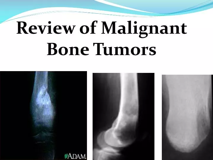
Review of Malignant Bone Tumors. Bone Tumors . Less than 1% of all tumors Rarely seen in podiatry: <2% of bone tumors occur in the foot/ankle 4x more likely to be benign vs. malignant. PATTERNS OF OSSEOUS DESTRUCTION. Evaluating lytic processes

E N D
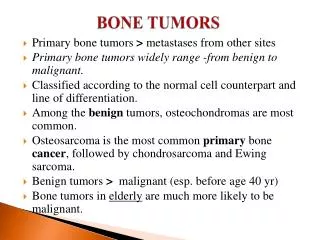
Bone Tumors • Less than 1% of all tumors • Rarely seen in podiatry: <2% of bone tumors occur in the foot/ankle • 4x more likely to be benign vs. malignant
PATTERNS OF OSSEOUS DESTRUCTION • Evaluating lytic processes • Can help identify lesion or create DDX (along with location) • Reflects aggressiveness of the lesion • Geographic • Moth-Eaten • Permeative
PATTERNS OF OSSEOUS DESTRUCTION • GEOGRAPHIC • Least aggressive • Indicative of a slow growing benign lesion • Well defined lesions, with narrow zone of transition from normal to abnormal bone
PATTERNS OF OSSEOUS DESTRUCTION • MOTH-EATEN • More aggressive, multiple small holes of internal lysis varying from 2 mm-5 mm • Seen around cortical bone, around edges of tumor • Wider zone of transition between normal and abnormal bone • Faster growing lesions, can be benign or malignant
PATTERNS OF OSSEOUS DESTRUCTION • PERMEATIVE • Most aggressive, rapidly growing, indicates malignancy • Countless number of small-caliber, <1.0 mm, holes of destruction in bone • Poorly defined lesions, with wide zone of transition, difficult to distinguish normal from abnormal bone.
SIGNS OF MALIGNANCY/PERIOSTEAL RESPONSE • SUNBURST • Rays of periosteal bone formation, separated by spaces containing blood vessels, “rays” are called Sharpey’s Fibers extending off of periosteum • Usually seen in Ewing’s sarcoma and Osteosarcoma
SIGNS OF MALIGNANCY/PERIOSTEAL RESPONSE • HAIR ON END • Similar to sunburst, but rays of periosteal new bone project parallel to each other, perpendicular to bone • Most malignant tumors
SIGNS OF MALIGNANCY/PERIOSTEAL RESPONSE • ONION SKIN • Production of multiple layers of new periosteal bone • Lesions growing unevenly give periosteum time to lay down new bone
SIGNS OF MALIGNANCY/PERIOSTEAL RESPONSE • CODMAN’S TRIANGLE • Usually indicates extremely aggressive tumor, lesion breaks through cortex and is then pushing into periosteum forming a triangle COMPLEX REACTIONS: Combination or multiple signs of periosteal response incicates most aggressive lesions, and increased chance of malignancy
OSTEOGENIC SARCOMA(OSTEOSARCOMA) • Bone forming malignant tumor, very aggressive • Most common malignant bone tumor • Young males 10-25 during rapid growth spurts, or adults >40 with preexisting condition (Paget’s) • Most common location: metaphysis of distal femur and proximal tibia in the knee region.
OSTEOGENIC SARCOMA(OSTEOSARCOMA) • Rapid progression of pain, swelling, fever • Elevated alkaline phosphatase , 2-3 times normal • Can be osteolytic or sclerotic so appearance can vary • Usually see a sunburst pattern or codman’s triangle as tumor expands and penetrates cortex • Treatment • Biopsy for DX, chest ct-whole body scan to rule out metastases, pre-op and post-op chemotherapy, • Prognosis is poor with 5 year survival rate 50%
CHONDROSARCOMA • Cartilage forming malignant tumor • Usually affects older adults, >40 years old, M>F • Most commonly seen in metaphyseal regions- femur, then calcaneus • Destructive, osteolytic lesion with some areas of calcification
CHONDROSARCOMA • CT scan, tumor usually sends metastases to lungs • Can arise from pre-exisiting benign tumor of cartilage • Slowly progressing pain • Usually results in amputation
EWING’S SARCOMA • Highly malignant tumor of unknown origin, usually located in the metadiaphysis region (femur, tibia, humerus, pelvis) • Range of 2-80 years old, however usually found in young children, 3:2 male to female ration, Caucasians >>African Americans
EWING’S SARCOMA • Pain, swelling for weeks to months with erythema and warmth over localized area • Intermittent fevers with leukocytosis, anemia, elevated ESR, and possible weight loss • Radiographically: Osteolytic lesions with moth-eaten appearance extending into soft tissue, onion skin periosteal reaction due to splitting and thickening of the cortex by the tumor, can be permeative in appearance-more aggressive.
EWING’S SARCOMA • Pathological fracture common • MRI needed to evaluate extent of soft tissue involvement • Treament • Surgical resection • Radiation Multi-drug and pre and post-op chemotherapy Metastasis to lungs and lymph nodes can occur • Leads to a poor prognosis
FIBROSARCOMA • Malignanct connective tissue tumor • Similar characterisitics to osteosarcomas, but produce fibrous tumor cells as opposed to bone tumor cells. • M=F, usually greater than 40 years old • Typically in metaphysis of femur or tibia
FIBROSARCOMA • Symptoms include: pain, swelling, limited motion, possible pathologic fracture • Radiographically: osteolytic with variable pattern of bone destruction, usually no associated periostitis • Aggressive tumor that has tendency to reoccur • Treatment: radiation, chemotherapy, surgical resection
MULTIPLE MYELOMA • Most common malignant bone tumor, however it is often considered a marrow cell tumor within the bone, rather than an actual bone tumor • Malignancy beginning in plasma cells of bone marrow • Impairs function of plasma cells to produce antibodies • Body then produces immunoglobin-Bence Jones proteins, which are ineffective against infections -skeletal pain, renal failure, recurrent bacterial infections
MULTIPLE MYELOMA • Rare in childhood, most common 6-7th decade • Up to 25% can have normal radiographic appearance • Sharply circumscribed lytic lesions or diffuse demineralization, osteopenia, • Rarely involves foot, however if seen in foot is hallmark for widepread of the disease process. • Treatment: no cure exists • Primary options: • Chemotherapy • Bone marrow transplant
References • Rhee JH. Lewis RB. Murphey MD. Primary Osseous Tumors of the Foot and Ankle. MagnReson Imaging Clin N Am. 2008. Feb. 16 (1) 71-91. • Chou,LB. Ho. Tumors of the Foot and Ankle. Foot and Ankle Int. 2009. Sep: 30 (9) 836-841. • Degroot, Henry. Comprehensive Source for Bone Tumor Information. Bonetumor.org. 2009. • Osher, L. Shook, J. Lo, K. Bone Tumors of the Foot and Ankle. McGlamry’s Comprehensive Textbook of Foot and Ankle Surgery. 1369-1429. • Dolce, Mark. Tumors of Bone. Present Lecture Series • Joyce, Micheal. Primary Malignant Bone Tumors. The Merck Manual: Online Medical Library. April 2008. • Ozger H. Eralp L. Basaran M. Surgical Treatment of Malignant Tumors of the Foot and Ankle.Int J. Clin. Oncol. 2005 Apr. 10 (2) 127-132