Download
1 / 52
570 likes | 943 Views
Lumbar Vertebrae, Sacrum and Coccyx. Chapter 8. Lumbar AP. Facility Identification Correct Marker Placement No Preventable Artifacts Correct Film Size (14 x 17 lw). Lumbar AP. Density Controlled by mAs Overall the density is not too dark or too light Contrast Optimal kVp 75-85

E N D
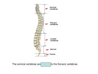
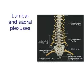
Lumbar Vertebrae, Sacrum and Coccyx Chapter 8
Lumbar AP • Facility Identification • Correct Marker Placement • No Preventable Artifacts • Correct Film Size (14 x 17 lw)
Lumbar AP • Density • Controlled by mAs • Overall the density is not too dark or too light • Contrast • Optimal kVp 75-85 • Bony trabecular patters and cortical outlines are well visualized • Vertebral bodies, pedicles, spinous processes, laminae, and pars interarticulari are demonstrated
Lumbar AP • True AP with no rotation • The spinous processes are in the midline of the vertebral body • The distance from the spinous process to each side of the vertebral body is equal • The sacrum and coccyx should be centered within the inlet pelvis and aligned with the symphysis
Lumbar AP • Detecting rotation • If the spinous processes are closer to one side of the vertebral body than the other • The side that the spinous process and lateral border of the vertebral body forms the greatest distance is the side rotated toward • Rotation can be either at the top (shoulders) or bottom (hips) or both
Lumbar AP • The Intervertebral disk spaces are open and the vertebral bodies are demonstrated without distortion (flex knees) • No foreshortening (hips or shoulders higher than the other) • The top and bottom of vertebral bodies are demonstrated if foreshortening is present
Lumbar AP • Align long axis of lumbar spine with film • Center L-4-5 to center of film
Lumbar Posterior Oblique • Facility Identification • Correct Marker Placement • No Preventable Artifacts • Correct Film Size (14 x 17 lw or 11 x 14 lw)
Lumbar Posterior Oblique • Density • Controlled by mAs • Overall the density is not too dark or too light • Contrast • Optimal kVp 75-85 • Bony trabecular patters and cortical outlines are well visualized • Vertebral bodies, pedicles, spinous processes, laminae, and pars interarticulari are demonstrated
Lumbar Posterior Oblique • Accurately rotated lumbar • Scotty dogs are visualized • Ear – superior articular process • Eye – pedicle (near the center of the body) • Neck – pars interarticularis • Feet – inferior articular process • Body - lamina
Lumbar Posterior Oblique • Degree of obliquity • Obliques too much • The “nose” of the Scotty dog is distorted (too short) and zygapophseal joint is closed • Not obliqued enough • The “eye” or pedicle of the Scotty dog appears too close to the lateral border of the spine
Lumbar Posterior Oblique • The long axis of the lumbar vertebral column is aligned with the long axis of the collimated field • The 3rd vertebra is in the center of the collimated field with T12 and 1st sacral segments visualized
Lumbar Lateral • Facility Identification • Correct Marker Placement • No Preventable Artifacts • Correct Film Size (14 x 17 lw)
Lumbar Lateral • Density • Controlled by mAs • Overall the density is not too dark or too light • Contrast • Optimal kVp 75-85 • Bony trabecular patters and cortical outlines are well visualized • Vertebral bodies, pedicles, spinous processes, laminae, and pars interarticulari are demonstrated
Lumbar Lateral • The lumbar vertebra are demonstrated in a true lateral position • The intervetebral foramina are clearly visualized • The spinous process are in profile • The right and left pedicles and the posterior surfaces of each vertebral body are superimposed
Lumbar Lateral • Detecting rotation • Evaluate the superimposition of the right and left posterior surfaces of the vertebral bodies
Lumbar Lateral • To detect anterior or posterior rotation • Locate 12th rib • Find magnified posterior rib • If magnified rib is anterior the patient is rotated then patient is rotated toward the anterior surface toward the film • If magnified rib is posterior the patient is rotated then the posterior surface is rotated closest to the film.
Lumbar Lateral • The intervetebral disk spaces are open • Vertebral bodies are demonstrated without distortion • Align the vertebral column parallel to the table top
Lumbar L5 – S1 Spot • Facility Identification • Correct Marker Placement • No Preventable Artifacts • Correct Film Size (8 x 10 lw)
Lumbar L5 – S1 Spot • Density • Controlled by mAs • Overall the density is not too dark or too light • Contrast • Optimal kVp 75-85 • Bony trabecular patters and cortical outlines are well visualized
Lumbar L5 – S1 Spot • True positioning • The 5th lumbar vertebra and sacrum are visualized • The intervetebral foramina are clearly visualized • Right and left pedicles are superimposed • Greater sciatic notches and pelvic wings are superimposed
Sacrum AP • Facility Identification • Correct Marker Placement • No Preventable Artifacts • Correct Film Size (10 x 12 lw)
Sacrum AP • The ischial spines are equally demonstrated and are aligned with the pelvic brim • Medial sacral crest and coccyx are aligned with the symphysis
Sacrum AP • Detecting rotation • Sacrum will rotate toward the side that is up • May also use pelvis criteria for detecting rotation
Sacrum AP • The 1-5 sacral segments are not foreshortened • The sacral foramina are equally spaced • The symphysis does not superimpose any portion of the sacrum
Sacrum Lateral • Facility Identification • Correct Marker Placement • No Preventable Artifacts • Correct Film Size (10 x 12 lw)
Sacrum Lateral • Density • Controlled by mAs • Overall the density is not too dark or too light • Contrast • Optimal kVp 75-85 • Bony trabecular patters and cortical outlines are well visualized
Sacrum Lateral • The median sacral crest is demonstrated in profile • The greater sciatic and pelvic wings are nearly superimposed • L5 – S1 disk space is open
Sacrum Lateral • The long axis of the sacrum is aligned with the film • The 3rd sacral segment is in the center of the film
Coccyx AP • Facility Identification • Correct Marker Placement • No Preventable Artifacts • Correct Film Size (8 x 10 lw)
Coccyx AP • Density • Controlled by mAs • Overall the density is not too dark or too light • Contrast • Optimal kVp 75-85 • Bony trabecular patters and cortical outlines are well visualized
Coccyx AP • The coccyx is aligned with the symphysis and is at equal distances from the lateral walls of the pelvic inlet
Coccyx AP • Detecting rotation • The coccyx will move in the same direction (toward) as the side that is up
Coccyx AP • The coccyx is in the center of the film • The symphysis, pelvic brim and 5th sacral segment are included on the film
Coccyx Lateral • Facility Identification • Correct Marker Placement • No Preventable Artifacts • Correct Film Size (8 x 10 lw)
Coccyx Lateral • Density • Controlled by mAs • Overall the density is not too dark or too light • Contrast • Optimal kVp 75-85 • Bony trabecular patters and cortical outlines are well visualized
Coccyx Lateral • The median sacral crest is demonstrated in profile • The greater sciatic notches are nearly superimposed
Coccyx lateral • Determining rotation • It is most common for the patient to be rotated anteriorly toward the table top if they do not have a sponge between the knees
Coccyx lateral • The first coccygeal vertebra is in the center of the film • S5 – the third coccygeal vertebra on film • Inferior median sacral crest are also on film