Download
1 / 4
60 likes | 259 Views
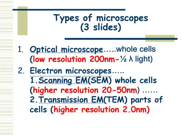
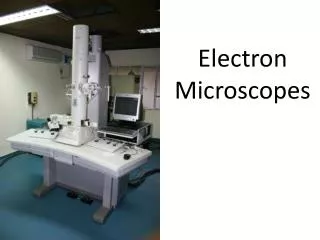
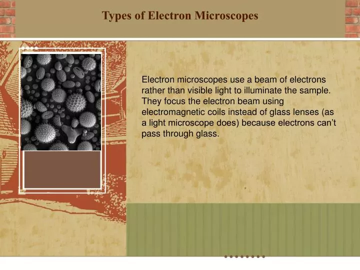
Types of Electron Microscopes. Electron microscopes use a beam of electrons rather than visible light to illuminate the sample. They focus the electron beam using electromagnetic coils instead of glass lenses (as a light microscope does) because electrons can’t pass through glass.

E N D
Types of Electron Microscopes Electron microscopes use a beam of electrons rather than visible light to illuminate the sample. They focus the electron beam using electromagnetic coils instead of glass lenses (as a light microscope does) because electrons can’t pass through glass.
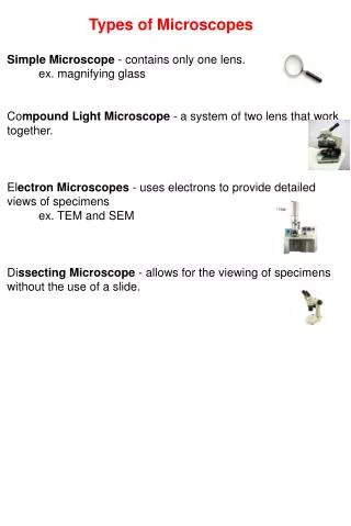
Scanning Electron Microscope (SEM) The scanning electron microscope (SEM) uses a focused beam of high-energy electrons to generate a variety of signals at the surface of solid specimens.
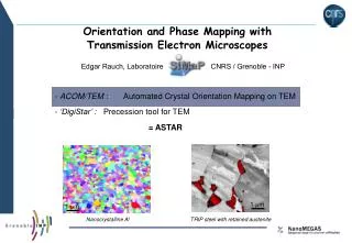
Transmission Electron Microscope (TEM) A "light source" at the top of the microscope emits the electrons that travel through vacuum in the column of the microscope. Instead of glass lenses focusing the light in the light microscope, the TEM uses electromagnetic lenses to focus the electrons into a very thin beam. The electron beam then travels through the specimen you want to study. Depending on the density of the material present, some of the electrons are scattered and disappear from the beam. At the bottom of the microscope the unscattered electrons hit a fluorescent screen, which gives rise to a "shadow image" of the specimen with its different parts displayed in varied darkness according to their density. The image can be studied directly by the operator or photographed with a camera.
Electron Microscope Pictures SEM versus TEM TEM SEM