Download
1 / 24
240 likes | 349 Views
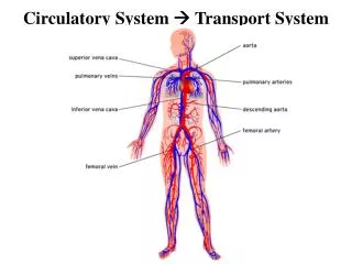
Topic 6: Human health & physiology. 6.2 The transport system. 6.2.1 Draw and label a diagram of the heart. Draw in the route of blood through the heart. The two sides of the heart (left and right) are separated by the septum Blood in the right side is deoxygenated ( oxygen-poor )

E N D
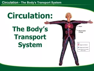
Topic 6: Human health & physiology 6.2 The transport system
6.2.1 Draw and label a diagram of the heart Draw in the route of blood through the heart
The two sides of the heart (left and right) are separated by the septum • Blood in the right side is deoxygenated (oxygen-poor) • Blood in the left side is oxygenated (oxygen-rich) • The right side of the heart is less muscular as the blood is pumped to the nearby lungs • Blood is under less pressure • The left side of the heart is more muscular as the blood is pumped to the rest of the body • Blood is under higher pressure Veins Arteries Low High Low (exception is the pulmonary vein) High (exception is the pulmonary artery)
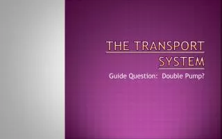
Blood is pumped around the body by the heart. It takes about 30 seconds for blood to go once around the body. Starting with the left side of the heart, what route does the blood follow to complete one circuit of the body?
body’s cells body’s cells The left side of the heart pumps oxygen-rich blood to the rest of the body. This blood supplies the body’s cells with oxygen. What gas does the blood then pick up from the body’s cells and where does the blood go next?
body’s cells body’s cells Blood picks up carbon dioxide from the body’s cells. This oxygen-poor blood then travels back to the right side of the heart. The oxygen-poor blood needs to lose the carbon dioxide and pick up more oxygen.
lungs lungs body’s cells body’s cells Next, the right side of the heart pumps oxygen-poor blood to the lungs. In the lungs the blood gets rid of the waste carbon dioxide and collects more oxygen.
body’s cells body’s cells The oxygen-rich blood then returns to the left side of the heart. This completes the blood’s journey around the body. lungs lungs
body’s cells body’s cells During one complete circuit of the body, blood passes through the heart twice. The heart has two jobs to do and so the circulatory system involves a double circulation. lungs lungs
blood vessels supply blood to muscle tissue 6.2.2 State that the coronary arteries supply the heart muscle with oxygen and nutrients The heart is full of blood but also needs its own blood supply so that the muscle can keep pumping. muscle tissue The blood vessels on the outside of the heart carry oxygen-rich blood to the heart muscle cells. Oxygen-poor blood is then carried away from these cells by outer blood vessels and back into the heart.
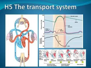
6.2.3 Explain the action of the heart in terms of collecting blood, pumping blood, and opening and closing of valves. • All the parts of the heart on either side, work together in a repeated sequence. • The two atria contract and relax; then the two ventricles contract and relax. • This is how blood moves through the heart and is pumped to the lungs and the body. • One complete sequence of contraction and relaxation is called a heartbeat.
When the heart muscles are relaxed – diastole • Blood flows from veins into atria • When the heart muscles contract – systole • Atria contract first to pump blood into ventricles (valve between atrium and ventricle opens) • Ventricles contract to pump blood into the arteries (forces valve to shut) Normal blood pressure: 120Systolic pressure 80 Diastolic pressure
6.2.4 Outline the control of the heartbeat in terms of myogenic muscle contraction, the role of the pacemaker, nerves, the medulla of the brain and adrenaline • The beating of the heart is due to myogenic muscle contraction... • This means that the myoctye (muscle cell) is the origin of the contraction and it is not controlled externally. • A region of myocytes, called the sinoatrial node (SA node) controls the rate of the heartbeat. • This region is also called the pacemaker. A wave of excitations is sent from the SA nose, causing the atria to contract. This excitation is conducted to the atrioventricular (AV) node, where it is passed through the nerves to the muscles of the ventricles, causing them to contract. The heart muscle is indefatigable. http://drugline.org/img/term/sa-node-13124_3.jpg
6.2.4 Outline the control of the heartbeat in terms of myogenic muscle contraction, the role of the pacemaker, nerves, the medulla of the brain and adrenaline (epinephrine) Heart rate can be controlled by the autonomic nervous system – the part of the nervous system that responds automatically to changes in body conditions. Where mycocardial contraction maintains the beating of the heart, we may need to speed up or slow down heart rate. When exercising, more CO2 is present in the blood. This is detected by chemoreceptors in the brain’s medulla oblongata, resulting in a nerve signal being sent to the SA node to speed up the heart rate. When CO2 levels fall, another nerve reduces heart rate. • The hormone adrenaline causes a rapid increase in heart rate in fight-or-flight reponses, preparing the body for action. • This effect can be mimicked by stimulant drugs.
6.2.5 Explain the relationship between the structure and function of the arteries, capillaries and veins.
thick outer wall thick inner layer of muscle and elastic fibres narrow central tube Arteries • Carry blood away from the heart • Except for the pulmonary artery, transport oxygenated blood • Artery walls are thick and elastic so they can stretch under the high pressure of blood • As the arteries stretch, they “pulse”
thin inner layer of muscle and elastic fibres wide central tube Veins • Carry blood to the heart • Except for the pulmonary vein, carry deoxygenated blood • Wider than arteries, with thinner walls • Blood under lower pressure • Valves prevent the back-flow of blood thin outer wall
artery vein Capillaries • Capillaries are the tiny blood vessels that carry a blood supply to and from the body’s cells. • Capillaries are the only blood vessels where substances can be exchanged between the blood and body cells.
6.2.6 State that blood is composed of plasma, erythrocytes, leucocytes and platelets • The liquid part of the circulatory system • Blood cells are suspended in plasma • An adult human has about 5.5L of blood in their body The four components of blood Plasma [55%] Erythrocytes (RBCs) [44.91%] Leucocytes (WBCs) and platelets [0.09%]
red blood cell white blood cell platelet • Blood plasma carries three types of blood cells. • They have different shapes and carry out different functions. Components of blood
Micrographs of blood Platelet Lymphocyte (WBC) Erythrocyte (RBC) Scanning electron micrograph of blood
6.2.7 State what is transported by the blood • nutrients, • oxygen, • carbon dioxide, • hormones, • antibodies, • urea, • heat http://www.webmd.com/heart/anatomy-picture-of-blood