Download
1 / 62
620 likes | 638 Views
Explore the different types of human tissues including epithelial, connective, muscle, and nervous tissues, their functions, and structures. Learn about glandular epithelium, types of glands, and connective tissue cells. Delve into the world of tissue biology with Meghna D. Punjabi.

E N D
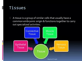
Tissues • A tissue is a group of similar cells that usually have a common embryonic origin & functions together to carry out specialized activities. Meghna.D.Punjabi
Epithelial Tissue: • Covers body surfaces; lines hollow organs,body cavities,and ducts;and forms glands. Meghna.D.Punjabi
Connective Tissue Protects & supports body and its organs, binds organs together, stores energy reserves as fats and provides immunity. Meghna.D.Punjabi
Muscle Tissue Responsible for movement and generation of force. Meghna.D.Punjabi
Nervous Tissue Initiates and transmits action potentials (Nerve impulses) that help coordinate body activities. Meghna.D.Punjabi
Epithelial Tissue • Cells are very closely packed & intracellular substance called matrix is minimal. • Cells lie on a basement membrane. Single layer of cells Several layer of cells Meghna.D.Punjabi
Simple Epithelium • Found on absorptive or secretory surfaces where single layer enhances this process. • Types named on the shape of cells & more active the tissue, the taller are the cells. Types of Simple epithelium Meghna.D.Punjabi
Squamous (Pavement) Epithelium • Single layer of flattened cells which fit closely like flat stones, forming a thin & smooth membrane. • Diffusion takes place freely through this thin, smooth, inactive lining of following structures:- heart, blood vessels, lymph vessels, alveoli of the lungs. Meghna.D.Punjabi
Cuboidal Epithelium • Cube shaped cells. • Forms the tubules of kidneys & some glands. • Involved in secretion, absorption & excretion. Meghna.D.Punjabi
Columnar Epithelium • Rectangular in shape. • Lining the organs of alimentary tract. • Some absorb the products of digestion & some secrete mucus. Meghna.D.Punjabi
Ciliated Epithelium • Columnar cells with hair like processes called cilia. • Cilia consists of microtubules. • Wave like movement of many cilia propels the contents of the tubes, which they line in one direction only. • Found lining the utrine tubes & respiratory passages. Meghna.D.Punjabi
Stratified Epithelia • Several layers of cells of various shapes. • Basement membranes absent. • Protects underlying structure from wear & tear. Keratinised Relaxed & Stretched Non-Keratinised Meghna.D.Punjabi
Stratified Epithelium: Meghna.D.Punjabi
Stratified Squamous Epithelium • Number of layers of cells • Deepest cells are columnar & as they grow towards the surface they become flattened & are then shed. • Non keratinised stratified epithelium (Wet surfaces) they are found in the conjunctiva of the eyes, lining of the mouth, pharynx, vagina, oesophagus. • Keratinised stratified epithelium (Dry surfaces) they are found in the skin, hair & nails, surface layer consists of dead epithelial cells to which the protein keratin has been added, this forms a tough, relatively water proof protective layer that prevents drying of the underlying live cells. Sub types Meghna.D.Punjabi
Transitional Epithelium: • Composed of several layers of pear-shaped cells. • Found: Lining urinary bladder. • It allows for stretching as bladder fills. Meghna.D.Punjabi
Glandular Epithelium: • Function: Secretion,accomplished by glandular cells that often lie in clusters deep to the covering and lining epithelium. • Gland may consists of one cell or a group of highly specialized epithelial cells that secrete into ducts,onto a surface or into the blood. • Production of such substances always require active work by the cells and results in an expenditure of energy. Meghna.D.Punjabi
Types of Glands: GLANDS EXOCRINE ENDOCRINE Meghna.D.Punjabi
Exocrine Glands: • Secrete their products into ducts(tubes) that empty at the surface of covering and lining epithelium or onto a free surface. • Product may be released at the skin surface or into lumen of hollow organs. • Secretions: Mucous,perspiration,oil,wax and digestive enzymes. • Ex: Sweat glands and Salivary glands. Meghna.D.Punjabi
Endocrine Glands: • Are ductless. • Secretory products enter the ECF and diffuse into blood. • Secretions always HORMONES,chemicals that regulate various physiological activities. • Ex: Pituitary,Thyroid snd Adrenal Glands. Meghna.D.Punjabi
Connective Tissue Cells forming are more widely separated from each other & intracellular substance (Matrix) is present in considerably larger amounts. Meghna.D.Punjabi
Cells of connective tissues Meghna.D.Punjabi
Fibroblasts • Large flat cells which produce collagen and elastic fibers and a matrix of extracellular material. • Particularly active in tissue repair (wound healing). Meghna.D.Punjabi
Fat Cells • Known as adipocytes these cells occur singly or in groups. • Vary in size and shape according to the amount of fat they contain. Meghna.D.Punjabi
Macrophages • Irregular shaped cells with granules in the cytoplasm. • Important part of the body’s defence mechanism as they are actively phagocytic,engulfing and digesting cell debris,bacteria and other foreign bodies. Meghna.D.Punjabi
Leukocytes • WBC’s found in small numbers in healthy connective tissue but migrate in significant number during infection when they play an important role in tissue defense. Meghna.D.Punjabi
Mast cells • Found in liver, spleen and around blood vessels. • Produce granules containing heparin, histamine etc. which are released when cells are damaged by diseases or injury. Meghna.D.Punjabi
Loose Connective Tissue • Found in every part of the body providing elasticity and tensile strength. • Connects and supports other tissues. • Example: Under the skin Between muscles, Supporting blood vessels and nerve cells, Alimentary canal, Glands supporting secretory cells. Meghna.D.Punjabi
Adipose Tissue Consists of fat cells Meghna.D.Punjabi
White Adipose Tissue 20 to 25% of body weight in adults. It is found supporting kidneys & the eyes, between muscle fibres & under the skin where it acts as a thermal insulator Meghna.D.Punjabi
Brown Adipose Tissue Found in newborn When brown tissue is metabolized produces less energy and considerably more heat than other fat , contributing to maintenance of body temperature. Meghna.D.Punjabi
Dense Connective Tissue Meghna.D.Punjabi
Fibrous tissue • Found forming ligaments, which binds bones together. • As an outer protective covering for bone called periostium. • Outer protective covering of some organs eg. kidneys,brain,lymph node. • Forming muscle sheaths called muscle facia which extends beyond the muscle to become the tendon that attaches muscle to bone. Meghna.D.Punjabi
Elastic Tissue • Considerable extension & recoil. • Few cells & the matrix consists mainly of masses of elastic fibres secreted by fibroblasts. • Found in organs where alteration of shape is required eg. Large blood vessels, epiglotis & outer ears. Meghna.D.Punjabi
Blood Meghna.D.Punjabi
Blood • Fluid connective tissue. • Consists of plasma & formed elements which include erythrocytes, leukocytes & thrombocytes. • Found within blood vessels (atreries, arterioles, capillaries, venules & veins.) • Transport oxygen & carbon dioxide; leukocytes carry on phagocytosis & are involved in allergic reactions & immunity, thrombocytes are essential for the clotting of blood. Meghna.D.Punjabi
Lymphoid Tissue • Found in blood & in the lymphoid tissue in the lymph nodes, spleen, palatine & pharyngeal tonsils, vermiform appendix and wall of large intestine. Meghna.D.Punjabi
Cartilage Firmer than any other connective tissue. Cells are chondrocytes and are less numerous & are reinforced by collagen& elastic fibres. Hyaline Cartilage Types Fibro Cartilage Elastic Cartilage Meghna.D.Punjabi
Hyaline Cartilage • Appears as a smooth bluish-white tissue. • Chondrocytes are in small groups within cell nests and the matrix solid and smooth. • Found: • On the surfaces of the parts of bones that form joints. • Forming the costal cartilages,which attach ribs to sternum. • Forming parts of larynx,trachea and bronchi. Meghna.D.Punjabi
Fibro Cartilage • Tough, slightly flexible tissue. • Found as follows:- • As pads between the bodies of the vertebrae called the intervertebral discs. • Between the articulating surfaces of the bones of the knee joint called semilunar cartilages. • On the rim of the bony sockets of the hip & shoulder joints deepening cavities without restricting movements. • As ligaments joining bones. Meghna.D.Punjabi
Elastic Cartilage Forms pinna or lobe of the ear, the epiglotis and part of the tunica media of blood vessel walls. Meghna.D.Punjabi
Bones Connective tissue with cells (Osteocytes) surrounded by a matrix of collagen fibres that is strengthened by inorganic salts especially calcium & phosphate. Meghna.D.Punjabi
Bones Types of Bones • Compact Bone: Solid or dense appearance • Compact bone consists of osteons that contain lamellae, lacunae, osteocytes, canaliculi & central canals. • Cancellous or spongy bone: Spongy of fine honeycomb appearance. • Spongy bones consists of thin plates called trabeculae. • Both compact & spongy bones comprise the various parts of bones of the body. • Support, protection, storage, houses blood forming tissue and serves as levers that act together with muscle tissue to provide movement. Meghna.D.Punjabi
Muscle tissue Meghna.D.Punjabi
Skeletal Tissue: Meghna.D.Punjabi
Skeletal Muscle Tissue: • Description: Cylindrical, striated fibres with many peripheral nuclei, voluntary control. • Location: Usually attached bones. • Function: Motion, posture, heat production. Meghna.D.Punjabi
Smooth Muscle Tissue: Meghna.D.Punjabi
Smooth Muscle Tissue: • Description: Spindle-shaped, nonstriated fibres with one centrally located nucleus; usually involuntary control. • Location: Walls of hollow internal structures such as blood vessels, airways to the lungs, stomach, intestines, gall bladder, and urinary bladder. • Function: Motion (constriction of blood vessels and airways, propulsion of foods through gastrointestinal tract; contraction of urinary bladder and gall bladder). Meghna.D.Punjabi
Cardiac Muscle Tissue: Meghna.D.Punjabi
Cardiac Muscle Tissue: • Description: Branched cylindrical, striated fibres with one or two centrally located nuclei, contains intercalated discs , mainly involuntary control. • Location: Heart Wall. • Function: Pumps blood to all parts of the body. Meghna.D.Punjabi
Nervous Tissue: Meghna.D.Punjabi


