Download
1 / 35
350 likes | 377 Views
Tissues. -four primary tissue types:. 1. Epithelial. 2. Connective. 3. Muscle. 4. Neural. provides support. binds structures together. fills cavities. produces blood. protects organs. Connective Tissue. Connective Tissue. 2 components: matrix + cells.

E N D
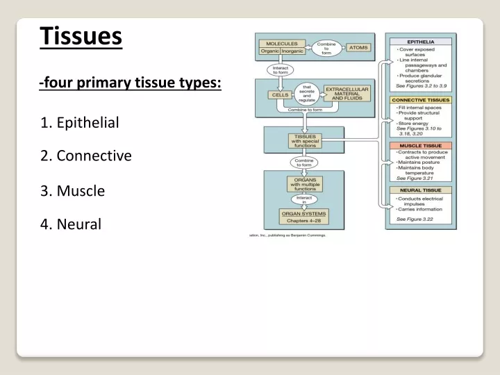
Tissues -four primary tissue types: 1. Epithelial 2. Connective 3. Muscle 4. Neural
provides support • binds structures together • fills cavities • produces blood • protects organs Connective Tissue
Connective Tissue • 2 components: matrix + cells -matrix:non-cellular support material -comprised of extracellular protein fibers – mainly collagens e.g. 1. collagen fibers (white) – collagen type I 2. elastic fibers (yellow) 3. reticular fibers – collagen type III 4. fibronectin -plus a ground substance = water + hyaluronan (sugar), proteoglycans and glycoproteins -cells: secrete the matrix -some have become very specialized and make a very specialized matrix
Connective Tissue • Loose – areolar CT, adipose & reticular • Dense – dense (regular, irregular), elastic • Supportive – bone & cartilage • Fluid – blood
Types: 1. Areolar 2. Dense – regular and irregular 3. Adipose 4. Cartilage 5. Bone 6. Blood
C H A P T E R F I V E THE SKELETAL SYSTEM: OSSEOUS TISSUE AND SKELETAL STRUCTURE Part A
Bones of the skeleton • Cartilage • Ligaments • Stabilize and connect bones Skeletal System
Structural support • framework for attachment • Storage of minerals and lipids • calcium bone salts (1-2 kg: 98% of it in bone) • Blood cell production, triglyceride storage • triglycerides stored in the yellow marrow • RBCs, WBCs, platelets in red bone marrow • Protection of delicate tissues and organs • ribs - heart and lungs; skull - brain; pelvis - digestive and reproductive • Leverage: change the magnitude and forces generated by skeletal muscles Functions of the Skeletal System
Supportive connective tissue • Solid extracellular matrix • framework of protein fibers: collagen type I • other specialized proteins: osteocalcin, osteonectin, osteopontin • ground substance – sugar + water: e.g. hyaluronan • calcium phosphate crystals: hydroxyapatite Osseous Tissue
Supportive connective tissue • Cells: osteoblasts, osteocytes, OPCs • cells only contribute total of 2% total bone tissue volume Osseous Tissue
Bone matrix is contains a large amount of hydroxyapatite crystals (calcium phosphate) Ca10(PO4)6(OH)2 • can withstand compression very well • 2/3 weight of bone • remaining 1/3 is collagen fibers, proteins & other salts • collagen contributes flexibility Bone: Histological Organization
also known as osteogenesis • osteogenesis involves calcification • deposition of calcium salts within a tissue • e.g. deposition of hydroxyapatite crystals among the “scaffolding” of collagen fibers and sugars • mechanism?? • controlled by the cells of bone Bone formation
Mature bone cells • maintain and monitor the matrix of bone • completely surrounded by hard bone matrix • directs the deposition of calcium within the bone matrix • do NOT undergo cell division Osteocytes
Immature bone-forming cells • cuboidal in shape • found on the inner and outer surfaces of bone • secrete the non-mineralized extracellular matrix = osteoid matrix • collagen I fibers + ground matrix + specialized bone proteins • this matrix will eventually become surrounded by hydroxyapatite crystals (i.e. calcification) • then matures to form an osteocyte • do NOT divide Osteoblasts
derived from stem cells (found within the bone marrow) • differentiate into osteoblasts • undergo division • play a role in fracture repair & bone remodelling Osteoprogenitor Cells (OPCs)
1. formation of OPCs from stem cells within bone marrow 2. differentiation of OPCs into osteoblasts (OBs) 3. OBs begins to produce the collagenous osteoid matrix 4. Osteoid calcifies into hard bone – formation of HA crystals around protein fibers of the osteoid 5. OBs mature into osteocytes 6. Bone maintenance by osteocytes Bone formation
large cells derived from macrophages • move into the bone – carving out tunnels by degrading the bony matrix (osteolysis) • through the production of mineral dissolving acids + protein-degrading enzymes called proteases • the components of the digested bone are recycled into the bloodstream by the OC • regulates blood calcium levels Osteoclasts
-new bone is made by osteoblasts -destruction of old bone is by osteoclasts - there must be balance between new bone production and old bone destruction to maintain bone volume -controlled by estrogen hormone action in females (increases activity of OBs) -also controlled in males and females by the hormones parathyroid hormone (stimulates OCs) and calcitonin (inhibits OCs) -disruption in this balance leads to disease e.g. osteoclast activity exceeds OB - loss of bone = osteoporosis e.g. too high OB activity – bone spurs Bone Remodelling
high turnover rate • 1/5 of skeleton is remodelled every month • takes 4 months to replace the femur completely Bone Remodelling
organizational pattern of bone matrix can be described as: • 1)compact bone • 2) spongy bone Types of Bone
also known as dense bone • makes up the bulk of the shaft of a long bone and the outer layers of short, flat and irregular bones • arranged in units called osteons or Haversian systems • osteons run the length of the bone’s shaft in long bone Compact Bone
central canal is the site for blood vessels, lymphatic vessels and nerves • bony matrix is organized as concentric circles around a central canal or a Haversian canal • concentric circles of bone around the central canal = concentric lamellae • osteons are aligned along the • bone in the same direction as • stress • e.g. shaft of a long bone - parallel to the long axis Osteons
within the lamellae are • small spaces in the matrix • containing osteocytes = lacunae • lacunae are interconnected • by canaliculi • the canaliculi are connected to the central canal • areas between osteons are filled with interstitial lamellaethat also have lacunae and canaliculi • are fragments of older osteons that are being degraded • large rings that surround the entire bone = circumferential lamellae
this is what calcified compact bone looks like under the microscope Bone histology
same matrix as compact bone • BUT different arrangement of osteocytes, canaliculi and lamellae • does not contain osteons • consists oftrabeculae= irregular lattice of thin bone columns Spongy Bone
spongy bone makes up the majority of the irregular, flat and short bones of the body • e.g. hip, ribs, sternum, ends of long bones (femur) • also found at the ends of long bones • these bones are covered with a thin layer of compact bone and filled with spongy bone Spongy Bone
the trabeculae of spongy bone are made up of lamellae with lacunae containing osteocytes and canaliculi • but what are trabeculae missing? • spaces between trabeculae are filled with red marrow • spongy bone is the site of hematopoiesis in adults (blood cell formation)
spongy bone is lighter - reduces overall weight of the bone • higher level of remodelling in spongy bone vs. compact (greater number of OBs and OCs within this type of bone) • stronger than compact bone - the trabeculae are arranged along lines of stress – highly resistant to breakage Spongy. vs. Compact Bone
Classification of bones • Based on anatomical classification • Long bones = greater length than width • Short bones = cube-shaped, spongy bone except at surface
Classification of bones • Flat bones = two parallel plates of compact bone sandwiching spongy bone layer
Classification of bones • Irregular bones = cannot be grouped • Sesamoid bones = develop in tendons where there is considerable friction, tension and stress • Sutural bones = located within joints between cranial bones
Anatomy of a Long Bone 1) diaphysis: shaft of the bone 2) epiphysis: distal & proximal ends of bone 3) metaphysis: region where the diaphysis joins the epiphysis -in a growing bone it includes the epiphyseal growth plate (hyaline cartilage) 4) articular cartilage: thin layer of hyaline cartilage covering the epiphysis -reduces friction during joint movement 5) periosteum: dense irregular connective tissue that covers the outside of the bone where not covered with articular cartilage -site of OPCs for bone formation 6) medullary cavity: or marrow cavity -space within diaphysis - contains yellow marrow 7) endosteum: thin membrane that lines the medullary cavity of long bones -single layer of bone forming OPCs
Periosteum and Endosteum • all bones are covered by periosteum (dense irregular connective tissue) • important for muscle attachment • source of OPCs • hollow chambers are lined with an endosteum
Circulatory Supply of a Bone -bones have a rich blood supply -blood vessels are abundant in bone portions containing red marrow -arteries + nerves enter the diaphysis and the epiphyses through holes called foramina (singular = foramen)