Download

1 / 40
410 likes | 1.09k Views
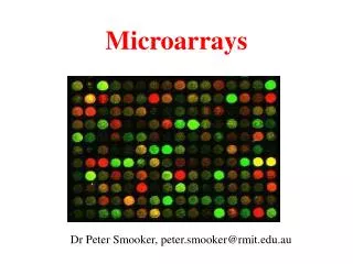
Antibody Microarrays. Merrill Birkner ph296~December 1, 2003. Antibody (Ab) Microarray. A complete microarray-based system for profiling protein expression in biological samples; used to compare two biological samples to measure the relative differences in protein expression.

E N D
Antibody Microarrays Merrill Birkner ph296~December 1, 2003
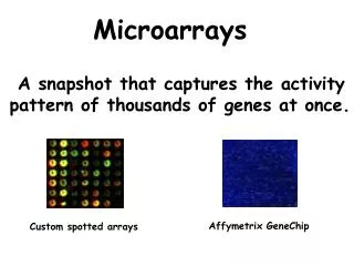
Antibody (Ab) Microarray • A complete microarray-based system for profiling protein expression in biological samples; used to compare two biological samples to measure the relative differences in protein expression. • The microarray consists of hundreds of monoclonal antibodies covalently bound in an ordered layout to a glass slide. • A protein which can be synthesized in pure form by a single clone (population) of cells. These antibodies can be made in large quantities and have a specific affinity for certain target molecules called antigens which can be found on the surface of cells and those that are malignant. • The array can be used as a means to correlate specific proteins with physiological or pathological process of interest, by comparing hundreds of proteins at a time. • It is used for toxicity testing, disease investigation, and drug discovery.
Antigens Antibodies • Antigens: • Molecules that stimulate the production of specific antibodies and combine specifically with the antibodies produced. Most antigens are foreign to the blood and other bodily fluids. • Antibodies: • Antibody proteins (immunoglobulins) are found in the gamma globulin class of plasma proteins. There are five main subclasses : IgG, IgA, IgM, IgD, and IgE. (ex. Most antibodies in serum are from the class IgG).
Antibody Structure • Consists of four interconnected polypeptide chains. Two heavy chains (H-chains) and joined to two shorter chains (L-chains). • These four chains are arranged in the form of a ‘Y’ ; with the stalk of the Y is called the “crystallizable fragment” and the top of the Y is known as the “antigen-binding fragment”.
Ab Array Procedure • Extraction of total cellular protein from biological samples of interest (eg. Serum samples). • Labeling of extracted protein with fluorescent dyes Cy5 and Cy3 (direct labeling, direct labeling with hapten tag, paired Ab sandwich assay). • Removal of unbound dye. • Incubation of labeled protein with the array. • Scanning of the array and the analysis of the results.
This procedure is a fluorescence-based analysis; covalently immobilized antibodies are used to capture fluorescently labeled antigens. • They do not measure absolute concentrations- instead they provide a relative measure of protein abundance [i.e. the abundance of protein in one sample as compared to another sample]. • As part of array development, all antibodies are printed and tested against their specific purified antigen (when available) and against cell lines and tissues samples (for quality control). • A reference pool is also used, and similar to the gene expression microarrays, a pool of equal aliquots from each sample to be measured is used, thus ensuring that all proteins from the samples are represented in the reference.
Direct Labeling • Direct Labeling with a hapten tag. • Paired Ab sandwich assays.
Direct Labeling (w/ hapten tag) • A convenient method to measure multiple proteins in a complex mixture. All proteins are labeled with either a fluorophore or a hapten tag such as biotin. • Advantages: • Only one captured antibody per target is required, as compared to the next method- easier to expand detection to new targets for which matched antibody pairs may not be available. • Can label different samples with different tags and to co-incubate the samples on the same arrays. • Disadvantages: • Potential for a high background: all proteins are labeled from the sample, including high concentration proteins such as albumin in serum; nonspecific binding or adsorption of these proteins to Ab could cause interference reduce detection sensitivity or data accuracy. • Potential for disruption of antibody-antigen interactions if the labeling reaction severely alters an antigen’s binding site.
Dual Antibody Sandwich • Antibodies spotted onto microarray substrates capture specific antigens, and a “cocktail” of detection antibodies, each antibody matched to one of the spotted antibodies, is incubated on the arrays. • Advantages: • Quantification of the bound detection antibodies provides a measure of each antigens abundance. • Sandwich assays are more sensitive than the direct labeling method because background is reduced through the specific detection of two antibodies instead of one. • Disadvantages: • The development and validation of assays measuring many targets in parallel is difficult because of the cross reactivity and precipitation when using many detection antibodies.
ELISA as a validation method • The Enzyme-Linked Immunoabsorbent Assay is serologic test used as a general screening tool for the detection of antibodies or antigens in a sample. ELISA technology links a measurable enzyme to either an antigen or antibody. • These tests are often used to validate the microarray results
Gene Expression vs. Ab Microarray • Gene expression, in most cases, does not necessarily correlate with changes in protein expression. • In cases when there is a correlation between mRNA and protein abundance, the correlation is often time shifted. • This time shift is likely to be different for each mRNA-protein pair. • With these arrays it is now possible to compare changes in gene expression with changes in protein expression using similar technologies. • There are also many reasons for merely studying protein abundance.
Ab Microarrays & Cancer Research • Information from protein profiling experiments may reveal associations between proteins or groups of proteins and disease states or experimental conditions. • Biomarkers in cancer are potentially valuable for early detection, staging of patients, classification of patients, or as surrogate markers for drug response. • These microarrays increase the number of proteins that can be conveniently measured, therefore taking advantage of the benefit of using combined markers in diagnostics.
Important in this field because there is a low volume requirement and the multiplex detection capability of microarrays make optimal use of precious clinical samples. • Work continues on the optimization of various aspects of the protocols, such as substrates for Ab attachment, the methods of Ab attachment, Ab buffers and concentrations, wash conditions, etc.
Antibody microarray profiling of human prostate cancer sera: Antibody screening and identification of potential biomarkers.Proteomics 2003, 3, 56-63. Miller, J., Zhou, H., Kwekel, J., Cavallo, R., Burke, J., Butler, E.B., Teh, B., Haab, B.
Background • Protein Biomarkers in the serum hold great promise for noninvasive disease detection and classification. • Ab & protein microarrays can have many applications including protein profiling of cancer tissue, autoimmune diagnostics, protein interaction screening, and Ab-based detection of multiple antigens. • Certain parts of the Ab microarray technology have not been perfected: • An optimized protein immobilization method is needed that retains native structure and reactivity and decreases nonspecific protein adsorption. • Ab can be immobilized by adsorption to poly-L-lysin membranes, by chemical cross–linking to derivatized glass surfaces. Hydrogels recently have also been introduced as a protein microarray substrate.
Another important issue is to create an efficient method of validating antibody performance in the microarray assay. • Previous work in the development of the antibody microarray methods made use of solutions of known target antigen concentrations to characterize antibody performance. • This is often very expensive and the antigens are often unavailable.
Goals 1. Compare two surfaces and antibody immobilization schemes: poly-L-lysine coated glass with a second photoreactive cross linking layer, & polyacrylamide-based hydrogels on glass. 2. Establish an efficient method to screen antibodies for those that are functional in the microarray assay. • Hypothesis: a statistical filter could identify antibody measurements that are consistent with specific and quantitatively accurate antigen binding. This hypothesis is tested by comparing microarray measurements to ELISA tests. 3. Demonstrate the use of this technology to screen serum samples for potential biomarkers, by analyzing the relative protein abundances in serum samples from prostate cancer patients and controls.
Serum samples • 33 males with prostate cancer ages 39-85 at the Methodist Hospital Houston, TX, USA, prior to commencement of radiotherapy. • PSA (prostate-specific antigen) concentration 2.5-335 ng/mL (from ELISA) median: 6.4 ng/mL • Histological grades of cancer tissue samples ranged from a Gleason combined scores of 6-9 • 20 serum samples taken from healthy males aged 30-69 • Normal PSA levels 0.2-3.2 ng/mL median: 0.85 ng/mL
Microarray preparation • The microarrays were deposited on two different types of substrates: the poly-L-lysine (HSBA) and the hydrogel [details found in paper]. • The serum samples and reference pool were diluted and mixed with the Cy5 or Cy3. • A reference pool is a pool of equal aliquots from each sample, thus ensuring that all proteins from the samples are represented in the reference.
Data analysis and statistics. • The local background in each color channel was subtracted from the signal at each antibody spot (spots with defects or no detectable signal removed). • The ratio of the net signal from the sample-specific channel to the net signal from the reference specific channel was calculated for each antibody spot; ratios from replicate antibody measurements in the same array were averaged. • The resulting ratios were multiplied by a normalization factor for each array (next slide) • Hierarchical clustering and visualization were performed using Cluster and Treeview. Ratios were log transformed & median centered. • Antibodies that did not have good measurements in at least 75% of the samples were removed from subsequent analysis. • The permutation t-test was calculated using the program Cluster Identification Tool.
Normalization Method • The resulting ratios were multiplied by a normalization factor for each array N, calculated by: N= (SIgG / μIgG)/RIgG SIgG = the ELISA-measured IgG concentration of the serum sample on that array. μIgG= the mean ELISA-measured IgG concentration of all of the samples. RIgG= the average ratio of the replicate anti-IgG antibody spots on a particular array.
Internal Normalization 4 slides per sample were created (label sample A with Cy3 and Cy5 and sample B with Cy3 and Cy5). Combine one of each of these samples and scan; determine the signal ratios of the 2 slides (Cy5/Cy3). With this method potential variability is eliminated because each protein sample labeled with each dye.
Results • Serum & control samples were analyzed using a two-color comparative fluorescence assay on microarrays containing 184 different antibodies spotted in quadruplicate. • 40 of the antibodies targeted 32 unique proteins that are typically found in the serum of healthy serum samples. • Another 13 antibodies targeted 9 proteins that have been detected in the serum of cancer patients, and the rest of the antibodies targeted normally intracellular proteins.
Goal #1 • The samples were repeated twice on the 2 types of slides (internal normalization); there were 4 microarray experiments per sample. • The hydrogel substrate generally produced a lower, more consistent background than the other surface. • The fluorescent signal from the Ab on the hydrogels had an average six-fold higher S/N ratio than the corresponding antibodies on the other surface. • The Ab showed measurable signal above background using the hydrogels (78 Ab) as compared to the other surface (23 Ab). • The hydrogel allowed weak detection from the greater number of Ab, reflecting decreased detection limits as a result of higher S/N ratio measurements.
Goal #2 • Define a statistical test that could filter the Ab measurement for those that are consistent with specific and accurate antigen binding. • They examined the overall variation in the reproducibility of Ab measurements from the 2 different surfaces after reverse labeling (mentioned before). • Developed an effective an efficient method to screen antibodies for those that function well in the microarray assay. • Using ELISA measurements as standards, they examined the ability of the statistical filter based on the correlation of the data from reversed labeling experiments to distinguish between reliable and unreliable microarray measurements.
First, in order to view patterns of similarity between sets of microarray measurements, average linkage hierarchical clustering is used. • There are 4 slides: hydrogel sample in red, hydrogel sample in green, HSBA sampled labels in red, and HSBA labeled in green. These were combined and clustered. • Each colored square represents one Ab measurement from one array. The color and intensity of each square represents the relative protein binding of the sample versus the reference. • Ab measurements that reproduce well between the different experiments are clustered together.
The correlation of measurements from replicate data sets as an initial screen to identify “reliable” antibodies. • The Pearson correlation of measurements between the reverse-labeled experiment set was calculated, both for hydrogel and HSBA. For both surfaces progressively fewer Ab exceeded the threshold as the threshold was increased. • In order to assess the degree to which the correlation parameter predicted specific and accurate antigen detection, microarray measurements from 7 of the Ab were compared to ELISA measurements for the corresponding antigens. • For these 7 antibodies a high inter-experiment set of correlations predicted a good agreement between the microarray and ELISA measurements. • They found no examples of Ab measurements that have high inter-experiment set correlation but poor agreement with ELISA measurements!
Goal #3 : Detection of biomarkers… • As a result of the previous analysis, only Ab that passed the stringent correlation threshold for inclusion were used in the following analysis. • They used a correlation threshold of 0.7, because the microarray measurements exceeding this threshold agreed well with the ELISA measurements. • In order to estimate the significance of the association between expression patterns and sample groups (cancer and normal) permutation t-tests were used. • Determines the statistical significance of each gene’s discrimination using a user defined segregation of samples.
Permutation t-test • Estimate the distribution of the t-test statistics under the null hypothesis by permutation of the sample labels. • The p-value pg is given as the fraction of permutations producing a test statistic that is at least as extreme as the observed one. It is the probability under the null hypothesis that the test statistic is at least as extreme as Tg. • Standard t-tests assume normally distributed data in each class and equal variance within classes. This test will be more accurate than the normal t-test for non-normal distributions and small samples.
When applied to the discrimination of cancer patients from the controls, CIT identified: • vWF, IgM, alpha-anti-chymotrypsin, Villin, and IgG with p-values below 0.01.
Hemoglobin was also discriminated but was found to be an artifact of hemolysis of controls. • None of these markers significantly correlated with PSA, and all varied independently of PSA. • IgM and IgG were lower and vWF higher in cancer patients & therefore similar to previous studies. • Since none of the proteins correlated with PSA, they could potentially bolster diagnostic accuracy if used in conjunction with PSA.
Remarks/Future work…. • Larger studies are needed to further examine the relationship between serum proteins and prostate cancer. • Further development in this technology will have significant utility in medical diagnostics as well as broader clinical and research application. • Using the D/S/A algorithm to analyze the data. (Data: Van Andel Inst. Michigan; Dr. Brian Haab)