Download
1 / 1
10 likes | 101 Views
Transcriptional and Proteomic R esponse to Iron L imitation in Pelagibacter ubique. Daniel P. Smith 1 , Joshua B. Kitner 2 , Angela D. Norbeck 3 , Therese R. Clauss 3 , Mary S. Lipton 3 , Mike S. Schwalbach 2 , Carrie D. Nicora 3 , Richard D. Smith 3 , & Stephen J. Giovannoni 2

E N D
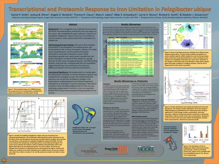
Transcriptional and Proteomic Response to Iron Limitation in Pelagibacter ubique Daniel P. Smith1, Joshua B. Kitner2, Angela D. Norbeck3, Therese R. Clauss3, Mary S. Lipton3, Mike S. Schwalbach2, Carrie D. Nicora3, Richard D. Smith3, & Stephen J. Giovannoni2 1Molecular and Cellular Biology Program, Oregon State University, Corvallis, OR 97331 • 2Department of Microbiology, Oregon State University, Corvallis, OR 97331 • 3Biological Science Division, Pacific Northwest National Laboratory, Richland, WA 99352 Abstract Background: Iron is recognized as an important micronutrient that limits microbial plankton productivity over vast regions of the oceans. We investigated the gene expression responses of Candidatus Pelagibacter ubique cultures to iron limitation in natural seawater media supplemented with a siderophore to chelate iron. Methodology/Principal Findings: Microarray data indicated transcription of the periplasmic iron binding protein sfuC increased by 16-fold, and iron transporter subunits, iron-sulfur center assembly genes, and the putative ferroxidase rubrerythrin transcripts increased to a lesser extent. Quantitative peptide mass spectrometry revealed that sfuC protein abundance increased 27-fold, despite an average decrease of 59% across the global proteome. Two RNA-binding proteins, CspE and CspL, correlated well with iron availability, suggesting that they may contribute to the observed differences between the transcriptome and proteome. Conclusions/Significance: We propose sfuC as a marker gene for indicating iron limitation in marine metatranscriptomic and metaproteomic ecological surveys. The marked proteome reduction was not directly correlated to changes in the transcriptome, implicating post-transcriptional regulatory mechanisms as modulators of protein expression. We propose a model in which the RNA-binding activity of CspE and CspL selectively enables protein synthesis of the iron acquisition protein SfuC during transient growth-limiting episodes of iron scarcity. Results: Microarrays The table below lists all 23 P. ubique mRNA transcripts that were at least 50 percent more abundant in the iron-limited cultures compared to the control cultures, 24 hours after addition of an iron-chelator. Shading indicates each gene’s location in Figure 3. This list is dominated by genes from the early (yellow) and late (green) iron stress clusters, indicating that P. ubique does not immediately modify its transcriptome for stationary phase when faced with iron scarcity. Results: Microarrays vs. Proteomics New dissolved iron supplied via aeolian deposition hslUV New dissolved iron supplied via upwelling/mixing suf Operon Figure 3: Genes transcribed during iron limitation were different from stationary phase genes. The four clusters indicate up-regulation of similar condition-specific mRNA. Symbols for each microarray sample (open circles) were manually positioned on a circle according to each sample’s iron availability and growth rate. Genes were “attracted” to the samples in which they were most abundant. Larger points indicate genes with larger condition-to-condition variation; a key for the 10 largest points in each cluster is provided. sfu Operon Total iron assimilation Summary P. ubique’s proteome bore little resemblance to its transcriptome. The genes listed in the above table, along with the unlisted lexA, recA, and mucA SOS-response genes also present in Figure 3’s late iron stress cluster varied greatly in mRNA abundance but were for the most part unchanging in protein abundance. Figure 4shows the sample-to-sample changes in protein abundances relative to the corresponding changes in mRNA abundances. Reaction to iron stress Iron-limitation had a marked impact on the overall proteome. Two days after addition of an iron-chelator, 181 of the 216 proteins were significantly (P≤0.05) less abundant in the iron-limited cultures relative to the control cultures (Figure 4, blue points). This abrupt decrease was independent of changes in mRNA abundance, showing that P. ubique’s general iron stress response is to reduce the size of its proteome via a post-transcriptional mechanism. We observed only one gene to be consistently up-regulated under iron limitation: sfuC. This protein localizes to the periplamic space where it associates with dissolved Fe3+ atoms and transports them to an ATP-driven permease. Due to the 16-fold increase in sfuC mRNA and 27-fold increase in SfuC protein under iron-limiting conditions, coupled with this organism’s high abundance in the oceans, we propose the use of sfuC as a biomarker for iron limitation in metatranscriptomic and metaproteomic environmental surveys. Adjusting to long-term limitation In Figure 4, the 44 purple points represent genes that significantly changed in either mRNA or protein abundance over time under iron-limiting conditions. The key observation here is that 68% of these genes showed an increase in protein abundance despite a decrease in mRNA abundance. RNA-binding proteins The abundances of the RNA-binding proteins CspE and CspL appear to correspond with iron availablility (Figure 5). Homologs of these proteins are able to selectively melt RNA secondary structures in Escherichia coli. We hypothesize that P. ubique uses CspE and/or CspL to regulate expression of proteins related to stress response; that they bind to specific mRNA secondary structures and melt them in order to facilitate ribosome attachment and allow the encoded genes to be translated into protein. Figure 1: These maps by Fung, et al. (2000) illustrate the varying marine iron concentrations resulting from aeolian dust deposition and deep water mixing. 10-7 10-6 10-5 10-410-3 mole Fe m-2 yr-1 Figure 4: Protein abundances were largely decoupled from transcript abundances. The change in protein abundance versus the change in mRNA abundance was plotted for all P. ubique genes that showed a significant (P ≤ 0.05) change in either measurement. Each color represents a different comparison between treatments or timepoints. Large ellipses indicate clusters of the same colored points. Histograms on the low end of each axis further define the distribution of points. Points represented by a diamond are discussed at left. Ferrichrome (below) sequesters dissolved iron in a complex that is unusable by P. ubique. Pelagibacter ubique cells, as imaged by cyro-electron tomography (Daniella Nicastro and Dick McIntosh, Univ. of Colorado). Figure 2: Growth of Candidatus Pelagibacter ubique cells was arrested by iron-sequestering siderophores. (A) Cell densities observed during a pilot experiment to test the effect of the two siderophores ferrichrome and deferoxamine mesylate salt at varying concentrations on the growth of Candidatus Pelagibacter ubique HTCC1062. The first arrow indicates the introduction of siderophore/iron as described by the legend. The second arrow indicates the delayed 1 µM iron additions parenthetically noted in the legend. (B) Cultures for harvesting were grown in six 20 L carboys. The first arrow indicates the introduction of siderophore/iron as described by the legend. Proteins and mRNA were analyzed on the dates indicated by the unfilled arrows: microarray samples were taken from cultures on days 17, 18, and 28; proteomic samples were taken on days 18 and 28. Figure 5: The abundance of two Ca. Pelagibacter ubique cold shock proteins, CspE and CspL, and the iron-binding protein SfuC, appear to be correlated with iron availability (p-value of .02, .08, and 3e-79, respectively). http://ncrr.pnl.gov http://www.mcb.oregonstate.edu/giovannoni http://moore.org/marine-micro.aspx