Download
1 / 23
520 likes | 1.16k Views
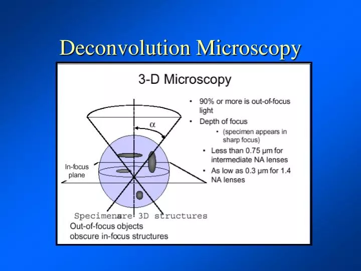
Deconvolution Microscopy. Deconvolution microscopy operates on the principle of a point spread function (PSF). As one moves away from focal plane in which an object lies the light from it will spread in a predictable manner.

E N D
Deconvolution microscopy operates on the principle of a point spread function (PSF). As one moves away from focal plane in which an object lies the light from it will spread in a predictable manner
The situation is analogous to what happens in an SEM, as we move away from the focal plane the beam spreads in a uniform and predictable manner
The point spread function must be measured for a given lens and a specific O.D. immersion oil
Much like a confocal a deconvolution system collects images in a Z-stack by repeatedly sampling the specimen at different focal planes
The components are much simpler consisting of a conventional fluorescence microscope, focus motor, CCD camera and computer with software.
Processing time for deconvolving images depends upon the size of the volume. A 32 by 32 by 32 volume takes 40 seconds to process on an Indigo2, 100 MHZ R4000 processor. Thus a 256 by 256 by 256 volume requires about 6 hours of processing time. Memory RequirementsMemory is an extremely important factor in determining how large a volume can be processed. To determine the required memory, three volumes must be present at one time: the original blurred volume, the 3D PSF and the deconvolved volume. This leads to the following table: Volume Size Required Memory 512 cubed 3.2 gigabytes 256 cubed 402 megabytes 128 cubed 50 megabytes 64 cubed 6.3 megabytes 32 cubed 785 kilobytes
The goal is to go from the 2D image stack to create a 3D confocal-like image
Removal of the out of focus portion of the image results in an in-focus section
Pollen Grain Muscle Cell Mitotic Cell
DeltaVison Deconvolution System: Commercial system with integrated CCD, stepper motor, and image processing station
DeltaVison Deconvolution System: Schizosaccharomyces
Advantages of Deconvolution over Confocal: Minimal equipment needed Microscope (with standard U.V. light source and filters) Focus Motor CCD Camera Computer and software No confocal or confocal aperture Light efficient since the entire image is collected at once (widefield)
Disdvantages of Deconvolution over Confocal: Processing time can be considerable (minutes to hours) Still restricted to monochromatic input (confocal can handle several different wavelengths simultaneously) Calculation of point spread function must be precise
TPEM = Two Photon Microscopy LSCM = Laser Scanning Confocal Microscopy DDM = Digital Deconvolution Microscopy
Deconvolution techniques can be used in: Widefield fluorescence microscopy Create in-focus Z-series Confocal and multiphoton microscopy Clean up and improve a Z stack Transmission electron microscopy Improve image contrast Scanning electron microscopy Achieve increased depth of field without decreasing resolution