Download
1 / 1
10 likes | 146 Views
a. b. c. MRI of experimental focal cerebral ischaemia in sheep. Annette Förschler 1) , Johannes Bolze 2) , Daniela Waldmin 3) , Uwe Gille 3) , Claus Zimmer 1) 1) Department of Neuroradiology, Leipzig University Hospital

E N D
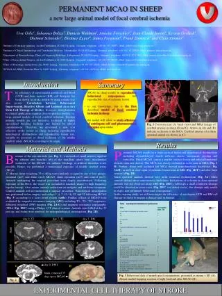
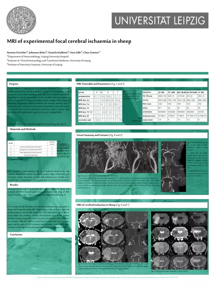
a b c MRI of experimental focal cerebral ischaemia in sheep Annette Förschler1), Johannes Bolze2), Daniela Waldmin3), Uwe Gille3), Claus Zimmer1) 1)Department of Neuroradiology, Leipzig University Hospital 2)Institute of Clinical Immunology and Transfusion Medicine, University of Leipzig 3)Institute of Veterinary Anatomy, University of Leipzig A new model of experimental focal cerebral ischaemia by permanent middle cerebral artery (MCA) occlusion in sheep was developed to study therapy for stroke with autologous stem cells from umbilical cord blood. Regarding the specific characteristic of rete mirabile epidurale rostrale in sheep we aimed to investigate the utility of time of flight (TOF) magnetic resonance angiography (MRA) to observe the vascular anatomy and to validate the MCA occlusion. Furthermore we intended to assess the extent and natural time course of ischaemic focal brain injury in sheep using functional and morphological magnetic resonance imaging (MRI). 13 Merino sheep were randomly assigned to one of four groups (fig. 1): Following exposure of the MCA branches, the vessels were occluded or touched (sham) by bipolar forceps. Controls did not undergo any surgical procedure. In 10 sheep 23 MRI sessions before and 2 to 46 days after onset of stroke (fig. 2) were performed using a 1,5T clinical MR scanner (fig. 3). Corrosion casts of the cerebral arteries of 3 sheep were prepared and compared to MRA. MRA visualised vessel anatomy (fig. 4) or occlusion distal to the rete mirabile. Anatomical variants concerning variant origin of the MCA and inconstant Arteria choroidea rostralis and communicans rostralis were revealed (fig. 5). Depending on the number of preserved MCA branches (0; 1; 2) significant (p<0,001) differences in lesion size (21±5,7; 13; 1,7±1,3 ml) could be found (fig. 6). In the sham operated animal no indications of ischaemia but a small contusion damage could be observed. Sheep with occluded left MCA showed space occupying lesions with drop of ADC values. From day 7 ADC values recovered and edema decreases (fig. 7). In our study for the first time focal cerebral ischaemia was generated in sheep and examined using MRI. Depending on the occlusion type the model produced reproducible lesion size. TOF-MRA proves to be able to clearly depict the anatomy, variants and occlusion type of the cerebral arteries in sheep comparable to the corrosion casts despite of the upstream rete mirabile. MRI with MRA is a useful tool to assess the extent of brain injury and the type of MCA occlusion and therefore is suitable for non-invasive monitoring of lesion development in stem cell therapy of stroke. Purpose MRI Timetable and Parameters (Fig. 2 and 3) Fig. 2: Timetable of the MRI sessions after the operation for each animal divided into groups. Fig 3: MRI parameters Materials and Methods Vessel Anatomy and Variants (Fig. 4 and 5) Fig. 1: Subgroups were build depending on the degree of MCA occlusion Fig. 5: MRA (MIP) of the rostral part of the circle of Willis displaying variant vessel anatomy: In variant (a) the middle cerebral artery arises from the distal part of the ICA. A clearly visible A. choroidea rostralis branches off proximally, running approximately parallel to the MCA. In variant (b) the MCA shows a more proximal origin. No second vessel can be identified. a b c a 1 3 16 2 5 15 10 6 11 14 12 b Fig. 4: Anatomy of the cerebral vessels in sheep (view from dorsal). (a) circumcision, (b) corrosion cast and (c) MR angiography: 1) A. cerebri rostralis, 2) A. communicans rostralis (inconstant), 3) A. cerebri media (MCA), 4) A. choroidea rostralis, 5) A. carotis interna (ICA, arising from 11), 6) A. communicans caudalis, 7) A. cerebri caudalis, 8) A. cerebelli rostralis, 9) A. basilaris, 10) Rami rostrales ad rete mirabile epidurale rostrale, 11) Rete mirabile epidurale rostrale, 12) Ramus caudalis ad rete mirabile epidurale rostrale, 13) A. cerebelli caudalis , 14) A. maxillaris, 15) A. buccalis, 16) A. ophthalmica externa. Results MRI of Cerebral Ischaemia in Sheep (Fig. 6 and 7) day 2 day 8 day 46 b1000 ADC T2* T2 Conclusion Fig. 6: Extend of the infarctions: (a) When preserving two of the branches of the MCA MRI shows only a small ischaemic lesion. (b) Focal ischaemia can be found, when one branch of the MCA remains in the MRA. (c) Complete middle cerebral artery occlusion with widespread infarction of the right hemisphere. Fig. 7: Time course of stroke in sheep (day 2/ day 8/ day 46). Authors correspondence: Annette Förschler, Universitätsklinikum Leipzig, Zentrum für Diagnostische Radiologie, Abteilung Neuroradiologie, Liebigstr. 20, 04103 Leipzig, Germany; E-Mail: annette.foerschler@medizin.uni-leipzig.de; Fon +493419717410