Download
1 / 1
10 likes | 209 Views
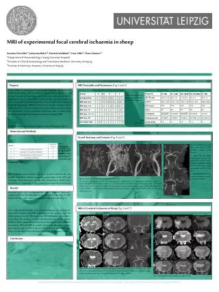
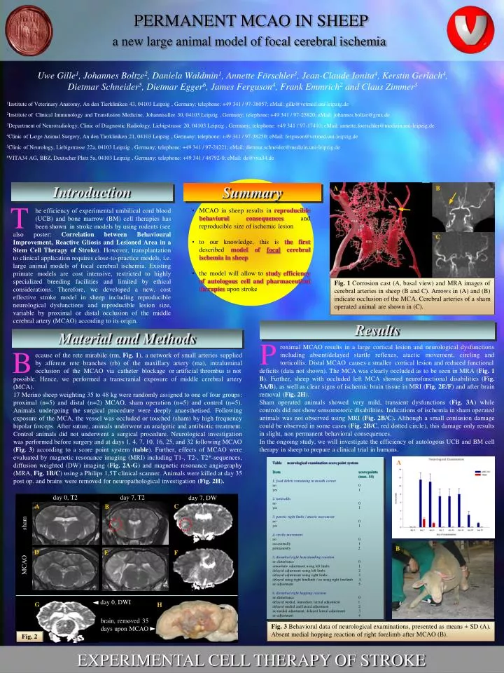
A. D. G. E. B. F. C. day 0, T2. day 7, T2. day 7, DW. sham. MCAO. H. day 0, DWI. brain, removed 35 days upon MCAO. a new large animal model of focal cerebral ischemia. PERMANENT MCAO IN SHEEP.

E N D
A D G E B F C day 0, T2 day 7, T2 day 7, DW sham MCAO H day 0, DWI brain, removed 35 days upon MCAO a new large animal model of focal cerebral ischemia PERMANENT MCAO IN SHEEP Uwe Gille1, Johannes Boltze2, Daniela Waldmin1, Annette Förschler3, Jean-Claude Ionita4, Kerstin Gerlach4, Dietmar Schneider5, Dietmar Egger6, James Ferguson4, Frank Emmrich2 and Claus Zimmer3 1Institute of Veterinary Anatomy, An den Tierkliniken 43, 04103 Leipzig , Germany; telephone: +49 341 / 97-38057; eMail: gille@vetmed.uni-leipzig.de 2Institute of Clinical Immunology and Transfusion Medicine, Johannisallee 30, 04103 Leipzig , Germany; telephone: +49 341 / 97-25820; eMail: johannes.boltze@gmx.de 3Department of Neuroradiology, Clinic of Diagnostic Radiology, Liebigstrasse 20, 04103 Leipzig , Germany; telephone: +49 341 / 97-17410; eMail: annette.foerschler@medizin.uni-leipzig.de 4Clinic of Large Animal Surgery, An den Tierkliniken 21, 04103 Leipzig , Germany; telephone: +49 341 / 97-38250; eMail: ferguson@vetmed.uni-leipzig.de 5Clinic of Neurology, Liebigstrasse 22a, 04103 Leipzig , Germany; telephone: +49 341 / 97-24221; eMail: dietmar.schneider@medizin.uni-leipzig.de 6VITA34 AG, BBZ, Deutscher Platz 5a, 04103 Leipzig , Germany; telephone: +49 341 / 48792-0; eMail: de@vita34.de A B Introduction Summary T • MCAO in sheep results in reproducible behavioral consequences and reproducible size of ischemic lesion • to our knowledge, this is thefirst described model of focal cerebral ischemia in sheep • the model will allow to study efficiency of autologous cell and pharmaceutical therapies upon stroke he efficiency of experimental umbilical cord blood (UCB) and bone marrow (BM) cell therapies has been shown in stroke models by using rodents (see also poster: Correlation between Behavioural Improvement, Reactive Gliosis and Lesioned Area in a Stem Cell Therapy of Stroke). However, transplantation to clinical application requires close-to-practice models, i.e. large animal models of focal cerebral ischemia. Existing primate models are cost intensive, restricted to highly specialized breeding facilities and limited by ethical considerations. Therefore, we developed a new, cost effective stroke model in sheep including reproducible neurological dysfunctions and reproducible lesion size, variable by proximal or distal occlusion of the middle cerebral artery (MCAO) according to its origin. C Fig. 1 Corrosion cast (A, basal view) and MRA images of cerebral arteries in sheep (B and C). Arrows in (A) and (B) indicate occlusion of the MCA. Cerebral arteries of a sham operated animal are shown in (C). Results Material and Methods P roximal MCAO results in a large cortical lesion and neurological dysfunctions including absent/delayed startle reflexes, atactic movement, circling and torticollis. Distal MCAO causes a smaller cortical lesion and reduced functional B ecause of the rete mirabile (rm, Fig. 1), a network of small arteries supplied by afferent rete branches (rb) of the maxillary artery (ma), intraluminal occlusion of the MCAO via catheter blockage or artificial thrombus is not deficits (data not shown). The MCA was clearly occluded as to be seen in MRA (Fig. 1 B). Further, sheep with occluded left MCA showed neurofunctional disabilities (Fig. 3A/B), as well as clear signs of ischemic brain tissue in MRI (Fig. 2E/F) and after brain removal (Fig. 2H). Sham operated animals showed very mild, transient dysfunctions (Fig. 3A) while controls did not show sensomotoric disabilities. Indications of ischemia in sham operated animals was not observed using MRI (Fig. 2B/C). Although a small contusion damage could be observed in some cases (Fig. 2B/C, red dotted circle), this damage only results in slight, non permanent behavioral consequences. In the ongoing study, we will investigate the efficiency of autologous UCB and BM cell therapy in sheep to prepare a clinical trial in humans. possible. Hence, we performed a transcranial exposure of middle cerebral artery (MCA). 17 Merino sheep weighting 35 to 48 kg were randomly assigned to one of four groups: proximal (n=5) and distal (n=2) MCAO, sham operation (n=5) and control (n=5). Animals undergoing the surgical procedure were deeply anaesthetised. Following exposure of the MCA, the vessel was occluded or touched (sham) by high frequency bipolar forceps. After suture, animals underwent an analgetic and antibiotic treatment. Control animals did not underwent a surgical procedure. Neurological investigation was performed before surgery and at days 1, 4, 7, 10, 16, 25, and 32 following MCAO (Fig. 3) according to a score point system (table). Further, effects of MCAO were evaluated by magnetic resonance imaging (MRI) including T1-, T2-, T2*-sequences, diffusion weighted (DW) imaging (Fig. 2A-G) and magnetic resonance angiography (MRA, Fig. 1B/C) using a Philips 1,5T clinical scanner. Animals were killed at day 35 post op. and brains were removed for neuropathological investigation (Fig. 2H). A Table neurological examination score point system Item score points (max. 14) 1. food debris remaining in mouth corner no 0 yes 1 2. torticollis no 0 yes 1 3. paretic right limbs / atactic movement no 0 yes 1 4. circlic movement no 0 occasionally 1 permanently 2 5. disturbed right hemistanding reaction no disturbance 0 immediate adjustment using left limbs 1 delayed adjustment using left limbs 2 delayed adjustment using right limbs 3 delayed using right hindlimb / no using right forelimb 4 no adjustment 5 6. disturbed right hopping reaction no disturbance 0 delayed medial, immediate lateral adjustment 1 delayed medial and lateral adjustment 2 no medial adjustment, delayed lateral adjustment 3 no adjustment 4 B Fig. 3 Behavioral data of neurological examinations, presented as means ± SD (A). Absent medial hopping reaction of right forelimb after MCAO (B). Fig. 2 EXPERIMENTAL CELL THERAPY OF STROKE