Download
1 / 45
450 likes | 646 Views
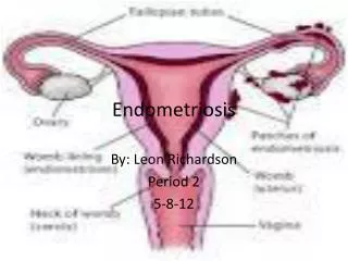
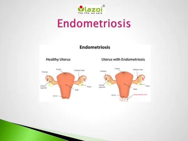
MRI IN DIAGNOSIS OF ENDOMETRIOSIS. INTRODUCTION . ENDOMETRIOSIS is a benign disorder characterised by proliferation of endometrial tissues outside the endometrial cavity. Most commonly between 30 -40 yrs 1/ 5 of all gynaecological cases . SITES. Ovary , Cul de sac (MC )

E N D
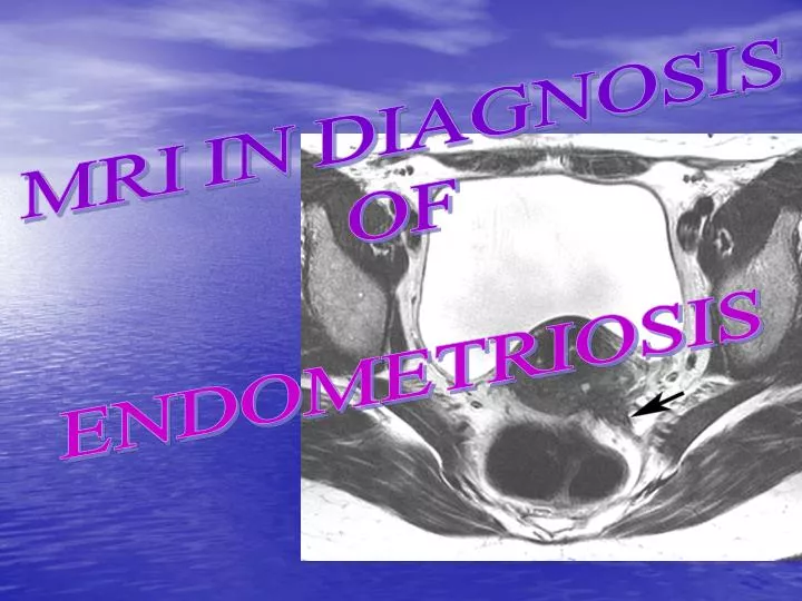
MRI IN DIAGNOSIS OF ENDOMETRIOSIS
INTRODUCTION • ENDOMETRIOSIS is a benign disorder characterised by proliferation of endometrial tissues outside the endometrial cavity. • Most commonly between 30 -40 yrs • 1/ 5 of all gynaecological cases
SITES • Ovary , Cul de sac (MC ) • Peritoneum covering the pelvic organs • GI and Urinary tract • Anterior abdominal wall • Extra peritoneal sites (Lungs and brain ) very rarely
CLINICAL PRESENTATIONS • Pelvic pain • Dysmenorrhea • Dyspareunia • Pain with defecation • Infertility • Multiple small nodules palpable along the uterosacral ligament during a PV examination
MRI • Technique based on magnetic properties of hydrogen nuclei • In the presence of a large magnetic field nuclear spin transition from ground state to the excited state can be induced • As the nuclei relax and return to the ground state energy is released in the form of electromagnetic radiation which is detected and processed into an image
Rectosigmoid colon endometriosis: sagittal T2-weighted MRI showing complete obliteration of the cul-de-sac due to intestinal endometriosis (white arrow) associated with endometrioma (arrowhead)
MIRM SEARCH • PUBMED • SCIENCEDIRECT • BIOSIS PREVIEW • SPRINGERLINK • PATENTSEARCH • GOOGLE SCHOLAR • BANDOLIER
LITERATURE REVIEW • INTRODUCTION • Deep pelvic endometriosis (DPE) is defined as the presence of endometrial implants, fibrosis and muscular hyperplasia below the peritoneum. DPE involves, in descending order of frequency, the uterosacral ligaments, the rectosigmoid colon, the vagina and the bladder . The exact incidence of DPE is unknown, but DPE is diagnosed in about one in five women with pelvic endometriosis.
PROCEDURE MATERIALS • MRI technique and analysisPatients fasted for at least 3 h before MRI and received an i.v. antispasmodic drug at the outset of the examination to decrease bowel peristalsis. MR images were acquired on a 1.5 T device. The protocol always included sagittal and axial fast spin-echo T2-weighted images and gradient echo T1 images with and without fat suppression, before and after injection of gadolinium. All sequences were acquired with anterior and posterior saturation bands placed anteriorly and posteriorly to eliminate the high signal of subcutaneous fat. Additional sequences could be performed, especially for suspected rectal involvement. The performance of the different sequences was not compared. • The MR images were analysed in real time by different radiologists
DISCUSSION Rectovaginal endometriosis: axial T2-weighted MRI passing through the lower limit of the cervix and showing an irregular hypointense endometriotic lesion of the rectovaginal septum (arrow)
Rectosigmoid colon endometriosis: sagittal T2-weighted MRI showing complete obliteration of the cul-de-sac due to intestinal endometriosis (white arrow) associated with endometrioma (arrowhead
Vaginal endometriosis: sagittal T1-weighted MRI showing hyperintense spots within the posterior vaginal fornix (arrow)
Rectovaginal septum.— Correlation between surgical and pathologic findings and MR imaging results was evaluated. All lesions of the rectovaginal septum that were found during surgery were resected. Lesions at this site of involvement were always accompanied by lesions of endometriosis at other posterior locations, such as USL (n = 10), vagina (n = 7), or rectosigmoid (n = 10) . Rectovaginal septum involvement . Posterior deep pelvic endometriosis was diagnosed at MR imaging in two patients with MR imaging results that were false-negative for rectovaginal involvement. Discrepancies between MR imaging and surgical findings were noted in regard to the precise anatomic location. The four false-positive results were observed in three patients with frozen pelvis (resection was not performed in two patients) and one patient in whom the precise extension of posterior involvement with endometriosis that was predicted with MR imaging differed from that seen during surgery. The sensitivity, specificity, positive and negative predictive values, and accuracy of MR imaging for the diagnosis of rectovaginal septum involvement compared with pathologic findings were 80% (eight of 10), 97.8% (181 of 185), 67% (eight of 12), 98.9% (181 of 183), and 96.9% (189 of 195), respectively. ( sample discussion , details in the report given )
TATS IT FR OUR TOPIC !! XIEXIE
GROUP MEMBERS • SARIKA PALEPU -12 • ARKAPAL BANDYOPADHYAY – 95 • ARNAB DUTTA -97 • ANUSHA MULKA -11 MBBS CHINA