Download
1 / 31
310 likes | 459 Views
Experimental Design and Analysis in Functional MRI. Thomas M. Talavage School of Electrical and Computer Engineering, Purdue University Department of Biomedical Engineering, Purdue University Department of Radiology, Indiana University School of Medicine. Overview.

E N D
Experimental Design and Analysis in Functional MRI Thomas M. Talavage School of Electrical and Computer Engineering, Purdue University Department of Biomedical Engineering, Purdue University Department of Radiology, Indiana University School of Medicine
Overview • Characterization of the fMRI Problem • Limitations Inherent in fMRI • Experimental Design and Analysis • The Future Thomas M. Talavage
The fMRI Problem • What we know: • Inputs • Time of each stimulus presentation • “Class” of each stimulus presented • Output • Measured signal as a function of space and time Thomas M. Talavage
The fMRI Problem • What we want to know: • The neural signal induced by an input signal • Function of space, X = (x,y,z) • Function of time, t • Difference in induced neural signals for similar (but not identical) inputs Thomas M. Talavage
The fMRI Problem • Definitions for a linear system interpretation • p(t): stimulus indicator function with range {0,1,…,L} • np(t)(X,t): neural signal induced by p(t) • rp(t)(X,t): hemodynamic response to np(t)(X,t) • s(X,t): observed image intensity • Combination of rp(t)(X,t) and physiologic and measurement noise terms, combined as h(X,t) Thomas M. Talavage
p(t) s(X,t) Human Subject in MRI System The fMRI Problem s(X,t) = + h(X,t) []* hvascular(X,t|p(t)) p(t) * hneuron(X,t|p(t)) np(t)(X,t) rp(t)(X,t) Thomas M. Talavage
The fMRI Problem • Solve for np(t)(X,t), given s(X,t) • Spatial localization of non-zero response to each p(t) • Temporal dynamics as function of p(t) • Observation is rp(t)(X,t) in noise, h(X,t) • Unknown parameterization of rp(t)(X,t) on p(t) • (Partially) uncharacterized noise, h(X,t) • “Level 2” Problem (Van Trees, 1968) • Goal: calculate np(t)(X,t) from estimate of rp(t)(X,t) Thomas M. Talavage
The fMRI “Problem” • But... Relation of rp(t)(X,t) to np(t)(X,t) is not well-defined Relation of rp(t)(X,t) to np(t)(X,t) may not be stationary Relation of rp(t)(X,t) to np(t)(X,t) may not be linear Thomas M. Talavage
Overview • Characterization of the fMRI Problem • Limitations Inherent in fMRI • Experimental Design and Analysis • The Future Thomas M. Talavage
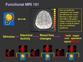
Limitations: Hemodynamic Response • hvascular(X,t|p(t)) not primarily a function of oxygen • Primary neural energy source is glucose • Neural activity depletes glucose reserves • Vascular response restores glucose reserves • Oxygen’s role in hvascular(X,t|p(t)) • Oxygen consumption a part of glucose usage • Vascular response maintains oxygen levels (epiphenomenon) • Oxygen consumption/delivery not balanced • Good: Leads to BOLD effect (Ogawa et al., 1990) • Bad:rp(t)(X,t) and hneuron(X,t|p(t)) not proportional? (Buxton et al., 1998) Thomas M. Talavage
Limitations: Measurement • Actual observation is discrete, s[X,nTR] • s(X,t) sampled at a period of TR, the repetition time • Temporal resolution O(100ms), more typically O(1s) • Neural activity: O(1ms) • Spatial resolution ~3mm • “Uncharacterized” noise, h(X,t) • Measurement (e.g., coil, drift) • Physiologic • Pseudo-deterministic: cardiac pulsation, respiration • Random: neural activity Thomas M. Talavage
Limitations: Measurement • Low contrast-to-noise ratio • BOLD effect signal (“contrast”) comparable to noise • Contrast-to-noise ratio affected by imaging process • Extent of imaged volume • 120-140 mm will cover whole brain; affects number of slices • Size of volume elements • Typically 3-4 mm in-plane; slice thickness generally 3+ mm • Sampling rate • More slices or higher spatial resolution increase sampling period • Slice acquisition takes 60-100 ms; usually sample every 2-3 s Thomas M. Talavage
Limitations: BOLD • Spatio-temporal properties • Temporal • “Late” onset relative to stimulus (~2s post-stimulus time, PST) • “Late” peak response for input impulse (~4s PST) • Slow decay (~10-15s PST) • Possible undershoot (~30s PST?) • Time-invariance? (e.g., Cauley et al., 2002) • Spatial • Stationary? (e.g., Saad et al., 2001) Thomas M. Talavage
Limitations: BOLD • Superposition of BOLD responses • Known non-linearity • Stimulus duration (e.g., Birn et al., 2001) • Simultaneous presentation of stimuli (e.g., Talavage and Edmister, 2004) • Function of which tissue is active? • Known limitation on dynamic range • Demonstrated via hypercapnia (Davis et al., 1998) • Response may be function of current state (Buxton et al., 1998) Thomas M. Talavage
Limitations: Subjects • Each human subject is unique • Performance • Multiple strategies for task performance? • Attention/Fatigue • “Rest” is typically not a resting state • Reaction to hostile (unnatural) environment • Confined space • Restricted movement • Acoustic noise Thomas M. Talavage
Overview • Characterization of the fMRI Problem • Limitations Inherent in fMRI • Experimental Design and Analysis • The Future Thomas M. Talavage
Restatement of fMRI Problem Solve for np(t)(X,t), given s(X,t) Solve for rp(t)(X,t), given s[X,nTR] Thomas M. Talavage
Overview of Design and Analysis • Answers to these questions guide choice of design and analysis procedures: • What change in strategy or taskis expected to produce a difference in the hemodynamic response, rp(t)(X,t)? • Where, as a function of X, will the differential activity in rp(t)(X,t) be observed? Thomas M. Talavage
Overview of Design and Analysis • Design of experiment should be based upon goal of analysis relative to rp(t)(X,t). • Two possible goals: • Maximize differences in rp(t)(X,t) across values of p(t) • Differences in amplitude as function of • Location, X = (x,y,z) • Time, t • Precisely estimate rp(t)(X,t) for various p(t) Thomas M. Talavage
Maximizing Differences • Goal: Detection of variation of amplitude at a given cortical location • Blocked Paradigm • Maximize rp(t)(X,t) by integration (step input) • Maximize measurements obtained during conditions A and B (NA and NB) to minimize error of means of measured signals, mA and mB. Thomas M. Talavage
Maximizing Differences • Goal: Detection of variation in time of response as a function of cortical location • Phase-Encode Paradigm • Monotonically vary p(t) with time • Identify monotonic latency variation (over X) in rp(t)(X,t) • “Continuous” mapping of response properties (e.g., receptive fields) Thomas M. Talavage
Estimating Responses • Goal: Estimation of response time-courses at a given position • Event-Related (ER) Paradigm (Buckner et al., 1996) • Temporally sparse distribution of “events” in p(t) • Maximize sampling rate (i.e., minimize TR) to densely sample rp(t)(X,t) between events • Average across events Thomas M. Talavage
Estimating Responses • Goal: Faster achievement of ER estimation of response time-courses at a given position • Rapid Presentation (RPER) Paradigm (Burock et al., 1998) • Uncorrelated sequence of “events” in p(t) • Maximize sampling rate (i.e., minimize nTR) to densely sample rp(t)(X,t) for all time, t • Average across events Thomas M. Talavage
Physiologically-Based Models • Goal: Augment detection and estimation through inclusion of a priori knowledge regarding BOLD response • Hemodynamic Response Functions (HRFs) • Constructed from physiological data • Optical imaging (e.g., Malonek and Grinvald, 1996) • Positron emission tomography (PET) (e.g., Rees et al., 1997) • fMRI (e.g., Mandeville et al., 1999) Thomas M. Talavage
Gamma variate model of Dale and Buckner (1997) Physiologically-Based Models • Model for hvascular(X,t|p(t)) • Single function: e.g., Gamma Variate (Dale and Buckner, 1997) • Multiple functions: e.g., Volterra kernel (Friston et al., 2000) Thomas M. Talavage
Significance of Responses • Most analyses assess activity in single voxel • “Activation” constructed by grouping voxels • Location • Proximity = subjective • Anatomy = objective(?) • Clustering • Statistical significance (valid?) • Orthogonal decomposition coefficients • Principal Component Analysis (e.g., McKeown et al., 1998) • Independent Component Analysis (e.g., Calhoun et al., 2001) Thomas M. Talavage
Overview • Characterization of the fMRI Problem • Limitations Inherent in fMRI • Experimental Design and Analysis • The Future Thomas M. Talavage
Future of Analysis • Goal: Capture four-dimensional (space+time) nature of response • Use priors to constrain spatio-temporal variation • s[X,nTR] • Descombes et al., 1998 • Rajapakse and Piyaratna, 2001 • HRF fit to s[X,nTR] • Purdon et al., 2001 • Solo et al., 2001 • Rao and Talavage, 2004 Thomas M. Talavage
Annual Meeting of the Organization for Human Brain Mapping (Budapest, Hungary) Future of Design • Goal: Measurement of np(t)(X,t) • Measurement of neuronal currents • Ideal to directly measure the quantity of interest • Small currents (microamp and smaller) • Short duration difficult to capture with MRI • The future is now...? • Bianciardi et al., 2004 • Kilner et al., 2004 • Park et al., 2004 Thomas M. Talavage
M Bianciardi et al., Org Hum Brain Mapp Annual Meeting, 2004. R Birn et al., Neuroimage 14:817-826, 2001. RL Buckner et al., PNAS USA 93:14302-14303, 1996. MA Burock et al., Neuroreport 9:3735-3739, 1998. RB Buxton et al., Magn Reson Med 39:702-708, 1998. VD Calhoun et al., Neuroimage 14:1080-1088, 2001. SF Cauley et al., IEEE EMBS Annual Meeting, 2002. AM Dale and RL Buckner, Hum Brain Mapp 5:329-340, 1997. TL Davis et al., PNAS USA 95:1834-1839, 1998. X Descombes et al., IEEE Trans Med Imaging 17:1028-1039, 1998. KJ Friston et al., Neuroimage 12:466-477, 2000. JM Kilner et al., Org Hum Brain Mapp Annual Meeting, 2004. SJ Kisner and TM Talavage, IEEE EMBS Annual Meeting,2004. D Malonek and A Grinvald, Science 272:551-554, 1996. JB Mandeville et al., Magn Reson Med 42:944-951, 1999. MJ McKeown et al., Hum Brain Mapp 6:160-188, 1998. S Ogawa et al., PNAS USA 87:9868:9872, 1990. TS Park et al., Org Hum Brain Mapp Annual Meeting, 2004. PL Purdon et al., Neuroimage 14:912-923, 2001. JC Rajapakse and J Piyaratna, IEEE Trans Biomed Eng 48:1186-1194, 2001. AA Rao and TM Talavage, IEEE EMBS Annual Meeting, 2004 G Rees et al., Neuroimage 6:270-278, 1997. ZS Saad et al., Hum Brain Mapp 13:74-93, 2001. V Solo et al., IEEE Trans Med Imaging 20:26-35, 2001. TM Talavage and WB Edmister, Hum Brain Mapp 22:216-228, 2004. TM Talavage et al., J Neurophysiol 91:1282-1296, 2004. HL Van Trees, Detection, Estimation, and Modulation Theory, Part I, 1968. RM Weisskoff et al., Proceedings Soc Magn Reson Med, 1992. Bibliography Thomas M. Talavage
The PhD Students S. Jordan Kisner Greg Tamer, Jr. Ashish Rao Sun Geun Kim Javier Gonzalez Castillo The MS Students Gilbert Tseng Ruwan Ranaweera Shrestha Basu Mallick Lejian Huang Chad Lau The Brave Undergraduate Ka Ki Ng Acknowledgments Purdue fMRI Research Group Thomas M. Talavage