Download
1 / 9
130 likes | 1.07k Views
Asymmetrical Corneal Topography in Map-Dot-Fingerprint Dystrophy Resembling Keratoconus. Arie L. Marcovich 1,2 , Ori Mahler 1,2 , Ayala Pollack 1 , Samuel Levinger 2. 1 Department of Ophthalmology, Kaplan Medical Center Rehovot, Israel

E N D
Asymmetrical Corneal Topography in Map-Dot-Fingerprint Dystrophy Resembling Keratoconus Arie L. Marcovich1,2, Ori Mahler1,2, Ayala Pollack1, Samuel Levinger2 1 Department of Ophthalmology, Kaplan Medical Center Rehovot, Israel 2 Enaim Laser Surgery, Tel Aviv & Jerusalem, Israel No Financial Interest
Background • Inmap-dot-fingerprint dystrophy, the corneal epithelium above an abnormal basement membrane, is not firmly attached to the underlying stroma, and may cause recurrent erosions. • Irregular epithelium can reduce vision Purpose To report 2 patients with map-dot-fingerprint corneal dystrophy with asymmetrical topographies that resembled keratoconus
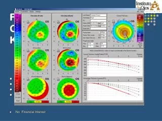
Patient 1 A 38 year-old female underwent corneal topography due to blurred vision in both eyes. Best corrected visual acuity was: OD 20/40 cc - 3.75 D OS 20/30 cc - 2.25 D Topography demonstrated superior asymmetry and the patient was referred to our cornea service with the diagnosis of keratoconus. EyeSys topography
Patient 1 Orbscan did not demonstrate posterior keratoconic changes.
Patient 1 On slit examination: In both corneas, signs of map-dot-fingerprint dystrophy were present. OD OS The patient was treated with sodium chloride 5% eye drops qid and lubricating ointment at night. Best corrected Visual acuity improved to 20/20 in both eyes with the same refraction.
Patient 2 A 41 year-old female complained of blurred vision in her right eye. She underwent extensive medical work-up including fluorescein angiography and MRI scan that were normal. Corneal topography was performed due to newly detected astigmatism in her right eye. 20/50 - 0.75 / + 3.25 X 160 20/20 without correction Topography showed inferior steepening in the right eye and a diagnosis of keratoconus was done. The patient was offered a surgical treatment with intracorneal rings.
Patient 2 Orbscan did not demonstrate posterior keratoconic changes. The patient was referred to our cornea service. On examination, signs of map-dot-fingerprint dystrophy were present in both corneas. The patient was treated with sodium chloride 5% eye drops qid and an ointment at night. Visual acuity did not improve, and the patient began to suffer from recurrent erosions in her right eye.
Patient 2 The patient underwent alcohol 20% assisted epithelial removal. Best corrected visual acuity improved to 20/20. Recurrent erosions did not recur in a one year follow up. Corneal topography demonstrated regular astigmatism. 20/50 - 0.75 / + 3.25 X 160 20/20 - 0.25 / - 0.50 X 138 OD inferior steepening misdiagnosed as keratoconus 10 months post alcohol delamination regular astigmatism on topography
Conclusions: • Map-dot-fingerprint dystrophy may cause blurring of vision and astigmatism due to epithelial irregularity, and induce topographic changes that may mimic keratoconus. • Asymmetry on topography may lead to misdiagnosis of keratoconus.