Download
1 / 12
130 likes | 313 Views
Coneal topography stability following 2.2 mm clear corneal phaco- emulsificaiton . Mohamed Hesham Aly , MD, FRCS Ed. ( Magrabi eye Hopital ) Bassel Atallah , OD ( Magrabi eye Hopital ).

E N D
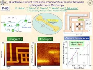
Coneal topography stability following 2.2 mm clear corneal phaco-emulsificaiton. Mohamed HeshamAly, MD, FRCS Ed. (Magrabi eye Hopital) BasselAtallah, OD (Magrabi eye Hopital) Neither the author nor the co- author has any financial interest in the items and products mentioned in this study.
Purpose Refractive surgery may be required post phaco-emulsification to correct residual astigmatism or miscalculated IOL power. The timing for the procedure should be decided after the corneal stability post phacoemulsification. A study was made to decide the time needed for the corneal topography to stabilize to be able to proceed for Excimer laser corrections.
Methods & results • 26 patients were followed up for 6 months post uneventful phaco-emulsification through 2.2 mm clear corneal temporal incision. All patients were done by the same surgeon. Infinity phaco machine (Alcon) was used using Ozil IP system. Corneal topography using Nidec OPD was done preoperatively, one day, one month, 2 months, 3 moths and 6 months post-operatively. • All patients were preoperatively assessed by routine ocular and systemic examinations. Assessment of corneal clarity, anterior chamber depth, pupil dilatation, fundus examination, all were performed as well as sonography and biometry. • Patients with corneal irregularities or disease were excluded from the study. Patients who needed wound suturing were also excluded. • The average astigmatic error was 0.538 preoperatively, increased to 0.959 one day post operatively, then reduced gradually until two months postoperatively to 0.51. The astigmatic error and corneal topography remained almost stable 2 months postoperatively till the end of the study (6months).
Conclusion Residual error corrections post phaco surgery, using Excimer laser correction, can be considered 2 months postoperatively. At this time most of the corneal topography changes should be stabilized.
Introduction • Cataracts are the leading cause of visual impairment and blindness in the world. Currently, phacoemulsification with intraocular lens (IOL) implantation is the most popular treatment for cataracts. With the improvement in surgical techniques and the development of new technology, the recovery of visual function has improved. • In recent years, phacoemulsification through a clear corneal tunnel incision has become increasingly popular due to ease of the technique, reduced length of surgery, little to no trauma to the conjunctiva, less surgically induced astigmatism, and quick rehabilitation of vision.
Introduction • Time needed for corneal topography stability should be evaluated if further procedure is needed to correct residual astigmatism. This is strongly recommended in patient asking for multifocal IOLs, miscalculated biometry and patient who is asking for refractive surgeries to get rid of distance glasses post phaco-emulsification surgeries. • The goal was to evaluate the time needed for the changes in corneal astigmatismand high order aberrations after surgery to stabilize after 2.2 mm temporal clear corneal incision surgery. • The outcomes of the study may assist the surgeon in selecting the proper time for the secondary corneal procedure to neutralize the resulting surgical astigmatic errors
PATIENTS AND METHODS A total of 26 patients (26 eyes) between the ages of 36 and 85 years (mean age: 60.52years) were followed up for 6 months. A 2.2-mm clear temporal (180 degree) corneal tunnel phacoemulsification/ IOL implantation was performed on the 26 eyes. Selection criteria included good general health, absence of corneal pathology during slit-lamp microscopy examination, no previous corneal or scleral surgery, absence of severe retinal pathology that could affect the infrared ray reflection from the macula during ocular wavefront aberration measurement, and no complications during or after surgery. An explanation of the study was given to all patients and informed consent obtained.
EXAMINATION Clinical examinations were conducted preoperatively and at 1 day, 1 weeks, 1 month, 3 months, and 6 monthsafter surgery. Clinical examination included best spectacle-corrected visual acuity (BSCVA) and uncorrectedvisual acuity (UCVA), manifest and cycloplegic refractions, intraocular pressure, and anterior and posteriorsegment evaluation. The corneal astigmatism and high order aberrations were measured using the NIDEKOPD-Scan aberrometer/topographer (NIDEK Co Ltd, Gamagori, Japan), which uses skiascopy-based ocularaberrometry using 1440 infrared points and placid disk corneal topography.7 The OPD-Station software(NIDEK Co Ltd) was used to isolate corneal aberration out to the sixth order.
SURGICAL TECHNIQUE Endocapsular phacoemulsification of the nucleus with Phacochop technique and cortical aspiration were performed using an Infinite(Alcon Laboratories). The anterior chamber and the capsular bag were refilled with Duovisc. A foldable IOL was inserted into the capsular bag using an injector cartridge system. The residual viscoelastic material was removed using bimanual irrigation/aspiration hand pieces. Balanced salt solution was injected through the paracentesis to maintain the anterior chamber. At the end of surgery, the wounds was checked and found to be watertight. All surgeries were completed without sutures. Postoperatively,all patients were treated with topical 0.1% Tobradex (Alcon Laboratories) and 0.1% Vigamos eye drops(Alcon Laboratories) every 2 hours for one week, then reduced gradually over one month period.
RESULTS Average pre-operative astigmatism was -0.935385 Dioptre. One day post-operatively, the average astigmatic error was -1.685384615 Dioptre. One week, one month, 2 months, 3 months, and 6 months post-operative astigmatic errors were -1.444615385, -1.335833333, -1.104615385, -1.033076923, and -1.02349804 respectively.
Results The average RMS pre-operatively was 0.538461538. The average RMS 1 day, 1 week, 1 month, 2 months, 3 months, and 6 months post-operatively were respectively: 1.006153846 0.763076923 0.534615385 0.653846154 0.661538462 0.658461538
DISCUSSION The optical quality of the pseudophakic eye is determined by the combination of corneal and internal aberrations generated by the IOL and those induced by surgery. Spherical aberration is determined by the corneal asphericity. In this study, corneal astigmatism and high order aberrations stability were evaluated over 6 months as measure of change in the cornea after cataract surgery. The need of the residual astigmatic and spherical aberration post-operatively corrections depends on the stabilization of these errors. Any premature correction may lead to augmentation or irregularities of these errors.