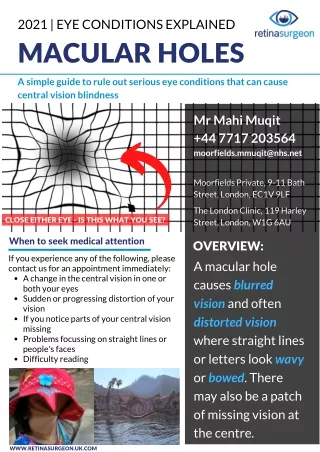
Download
1 / 30
350 likes | 755 Views
Macular Corneal Dystrophy. Matthew Kaufman, MD Ophthalmic Pathology CPC NP Fellow: Ken Clark, MD Attending: Charleen T. Chu, MD, PhD. Initial Presentation. 50 year-old male with a chief complaint of blurred vision and glare from both eyes Diagnosed with corneal dystrophy 6 years prior

E N D
Macular Corneal Dystrophy Matthew Kaufman, MD Ophthalmic Pathology CPC NP Fellow: Ken Clark, MD Attending: Charleen T. Chu, MD, PhD
Initial Presentation • 50 year-old male with a chief complaint of blurred vision and glare from both eyes • Diagnosed with corneal dystrophy 6 years prior • No significant past medical history • Visual Acuity: • 20/40 OD • 20/30 OS • Pupils, EOMS, Visual Fields, and IOPs Normal
Exam • Slit lamp exam revealed large nummular stromal opacities in the cornea with hazy intervening stroma • Opacities were more superficial centrally and more posterior peripherally • No epithelial staining with fluorescein
Pathology – Cornea OS (H&E) Epithelium Mucopolysaccharide deposits Stroma
H&E Mucopolysaccharide
Alcian Blue (pH 2.5) Mucopolysaccharide
Colloidal Iron Mucopolysaccaride
Descemet’s Membrane H&E Gutta
Diagnosis and Plan • Macular Corneal Dystrophy • Patient was offered phototherapeutic keratectomy (PTK) with plan for lamellar keratoplasty or penetrating keratoplasty if PTK was ineffective • Given minimal symptoms, patient chose to defer treatment
Two Years Later… • Patient complains of increased difficulty driving and reading • Reports significant glare symptoms • Visually acuity 20/50 OU • Proceeds with PTK OD • Post-op visual acuity 20/30 with improved symptoms
One year post-PTK • Patient feels vision worsening again • Difficulty reading, seeing a golf ball, and driving. Still having significant glare/haze. • Slit lamp exam shows the dystrophy affecting all levels of the corneal stroma • Patient elected to proceed with penetrating keratoplasty (PKP) in the left eye
Case • PKP performed in patient’s left eye • Pathology showed corneal stroma with glycosaminoglycan/mucopolysaccharide deposits • Stained positively for for alcian blue and colloidal iron • Guttata were present on Descemet’s membrane
Conclusion of Case • Nine months following PKP in the left eye, vision improved to 20/30 • Right eye vision remains 20/40 • Improvement in glare
Macular Corneal Dystrophy • Autosomal recessive inheritance • Mutation in chromosome 16q22 • Less common than lattice and granular dystrophies • Most prevalent in India, Saudi Arabia, Iceland, and parts of the USA • Corneas clear at birth • Clouding usually begins in the first decade • Earlier onset than lattice and granular • Progressive decrease in vision usually resulting in severe visual impairment
Clinical Appearance • Bilateral • Ill-defined gray-white stromal opacities with hazy surrounding stroma • As disease progresses, hazy areas of stroma merge leaving no intervening clear zones • Progressive extension through entire thickness of central and peripheral corneas • Endothelium and Descemet’smay be involved and show guttata • Rarely, recurrent erosions develop
Three Subtypes • Type I • No detectable keratan sulfate (KS) in the serum or cornea. Normal dermatan sulfate-proteoglycan (DS) • Type II • Normal ratio of KS to DS, but 30% lower production than normal with DS chains 40% shorter than normal • Type IA • No KS in the serum, but KS present in keratocytes
Workup • ELISA to measure sulfated keratan sulfate in preclinical forms or carriers
Histopathology • Intracytoplasmic inclusions in keratocytes and endothelial cells of acid mucopolysaccharide (glycosaminoglycans) • Stain positively for the following: • Alcian blue • Colloidal iron • Sometimes Periodic acid-Schiff • May have guttata of Descemet’s membrane
Classic Corneal Stromal Dystrophies • Macular Corneal Dystrophy • Lattice Corneal Dystrophy • Granular Corneal Dystrophy • Avelino Dystrophy
Lattice Corneal Dystrophy • Type I: TGFBI gene, mutation in locus 5q31 • Autosomal dominant • Clinical: • Glasslike branching lines in stroma • Pathology: • Amyloid deposits in anterior stroma extending to subepithelial area with possible disruption of epithelium • Stains with Congo red dye • Recurs in PKP more frequently than granular or macular
Lattice Dystrophy Congo red staining amyloid Apple-green birefringence under polarized light Source:odlarmed.com/?p=3684 Source: webeye.ophth.uiowa.edu
Granular Corneal Dystrophy • TGFBI gene, mutation in locus 5q31 • Autosomal dominant • Clinical: • Crumblike stromal opacities with intervening clear spaces • Pathology: • Hyaline rod-shaped deposits in stroma that can extend anteriorly through focal breaks in Bowman’s layer • Stains with Masson trichrome
Granular Dystrophy Source: odlarmed.com/?p=3680 Masson trichrome staining hyaline Source: webeye.ophth.uiowa.edu
Avellino Dystrophy • TGFBI gene, locus 5q31 • Autosomal dominant • Combination of lattice and granular dystrophies • Named for 4 early patients who traced their family to Italian province of Avellino • Clinically and pathologically shows granular and lattice lesions • Stains with both Masson trichrome and Congo red dye
Avellino Dystrophy Source: webeye.ophth.uiowa.edu
References Case ID: PHS11-34882 Clinical Approach to Corneal Dystrophies and Metabolic Disorders. In: Basic and clinical science course (BCSC) Section 8: External disease and cornea. San Francisco, CA. American Academy of Ophthalmology; 2009:319-20. Klintworth, GK. Corneal dystrophies. Orphanet J Rare Dis. 2009 Feb 23; 4:7. Review.