Download
1 / 27
280 likes | 401 Views
Introduction to Anatomy. Diane A. Young Brewbaker Technology Magnet High School. Anatomy. Anatomy – study of the structures of the body Physiology – study of the functions of these structures. Body Planes. VERTICAL PLANES: Vertical – up-and-down line at the right angle to the horizon

E N D
Introduction to Anatomy Diane A. Young Brewbaker Technology Magnet High School
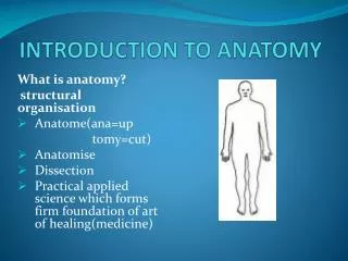
Anatomy • Anatomy – study of the structures of the body • Physiology – study of the functions of these structures
Body Planes VERTICAL PLANES: Vertical – up-and-down line at the right angle to the horizon Midsagittal plane (midline) – the vertical plane that divides the body, from top to bottom into equal left and right halves. Sagittal plane – any vertical plane parallel to the midline that divides the body into unequal left and right portions. Coronal plan (frontal) – any vertical plane, at right angles to the sagittal plane that divides the body into anterior (front) and posterior (back) portions.
Body Planes • Horizontal Planes: • Transverse plane (horizontal) – divides the body into superior (upper) and inferior (lower) portions • A transverse plane can be at the waist or any other level of the body
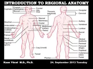
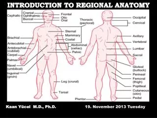
Body Directions • Body directions – relative location of the whole body or an organ is described through the use of pairs of contrasting body direction terms. • Ventral – the front or belly side of the body or organ • Anterior – situated in the front. Also, the forward part of an organ. • Superior – uppermost, above or toward the head • Cephalic – toward the head • Proximal – situated nearest the midline or beginning of a body structure • Medial – direction toward or nearer the midline
Body Directions, cont’d • Dorsal – to the back of the body or organ • Posterior – situated in the back or on the back part of an organ. • Inferior – lowermost, below, or toward the feet • Caudal – toward the lower part of the body • Distal – situated farthest from the midline or beginning of body structure • Lateral – the direction toward or nearer the side and away from the midline • Bilateral – relating to, or having, two sides
Examples • Anterior: • The mouth is anterior to the oral cavity. • The trachea (windpipe) is anterior to the cervical spine (vertebrae). • The skin of the palm is anterior to the wrist bones (carpals). • The patella is anterior to the knee joint. • Posterior: • The oral cavity is posterior to the mouth. • The cervical vertebrae is posterior to the trachea.
Examples cont’d • Superior: • The forehead is superior to the nose. • The head is superior to the neck. • The most superior portion of the femur is the femoral head. • The humerus is superior to the elbow joint. • The heart is superior to the diaphragm • Inferior: • The nose is inferior to the forehead. • The neck is inferior to the head
Major Body Cavities • Dorsal Cavity divided into two parts, protects the structures of the nervous system that coordinate the bodily function • Cranial cavity – located within the skull, protects the brain • Spinal cavity – located within the spinal column, protects the spinal cord
Major Body Cavities • Ventral Cavity divided into three parts, contains many of the body organs that maintain homeostasis. Homeostasis means maintaining a constant internal environment. • Thoracic cavity (chest cavity) – protects the heart and lungs. The diaphragm is a muscle that separates the thoracic and abdominal cavities. • Abdominal cavity – contains primarily the major organs of digestion. Referred to as the abdomen. • Pelvic cavity – the space formed by the pelvic (hip) bones. It contains primarily the organs of the reproductive and excretory systems. • There is no division between the abdominal and pelvic cavities. Together they may be referred to as the abdominopelvic cavity.
Division of the Abdomen • To make it easier to describe where an organ or a pain is located, the abdomen is divided into four imaginary quadrants. • Right upper quadrant (RUQ) • Left upper quadrant (LUG) • Right lower quadrant (RLQ) • Left Lower quadrant (LLQ)
Regions of the Thorax and Abdomen • Right and left hypochondriac regions. Hypochondriac means below the ribs. This term also means the individual with an abnormal and excessive concern about his/her health. • Epigastric Region – epigastric means above the stomach • Right and left lumbar region. Lumbar refers to the inward curve of the spine • Umbilical region-The term umbilical means referring to the umbilicus (belly button or naval). • Right and left iliac regions- Iliac refers to the hipbone. • Hypogastricregion -Hypogastric means below the stomach. The entire lower region of the abdomen is also referred to as the groin or inguinal area.
Peritoneum • Peritoneum is the membrane that protects and supports (suspends in place) the organs located in the abdominal cavity. • Parietal peritoneum – the outer layer of this membrane that lines the abdominal cavity • Visceral peritoneum – the inner layer of this membrane that surrounds the organs of the abdominal cavity. Visceral means relating to the internal organs (those enclosed within a cavity) especially the abdominal organs. • Mesentery – a layer of the peritoneum that suspends parts of the intestine within the abdominal cavity. • Retroperitoneal – means located behind the peritoneum of the abdominal cavity.
Peritoneum, cont’d • Peritonitis– inflammation of the peritoneum. • Ascites – an abnormal accumulation of clear or milky serous (watery) fluid in the peritoneal cavity.
Cytology • Cytology is the study of the formation, structure, and function of the cells. • Cell membrane – the structure that surrounds and protects the cell. A membrane is a thin layer of tissue that covers a surface, lines a cavity or divides a space or organ. • Cytoplasm is the material within the cell membrane that is not part of the nucleus. • Nucleus – is surrounded by the nuclear membrane. It is a structure within the cell that has two important functions: Controlling the activities and helps the cell divide. Cells are the basic structural units of the body that are specialized and grouped together to form the tissues and organs.
Tissue • The four main types of tissue are epithelial, connective, muscle and nerve • Epithelial tissue – form a protective covering for all of the internal and external surfaces of the body. • Epithelium – the specialized epithelial tissue that forms the epidermis of the skin and the surface layer of mucous membranes. • Endothelium – specialized epithelial tissue that lines the blood and lymph vessels, body cavities, glands and organs • Glands are made up of specialized epithelial tissues that are capable of producing secretions.
Tissue • Connective tissues support and connect organs and other body tissues • Adipose tissue, also known and fat, provides protective padding, insulation, support and acts as a nutrient reserve. • Loose connective tissue – surrounds various organs and supports both nerve cells and blood vessels. • Blood and lymph are liquid connective tissue • Bone and cartilage are dense connective tissue • We will discuss blood, lymph, bone and cartilage later
Tissue • Muscle tissue – contains cell material with the specialized ability to contract and relax • Nerve Tissue – contains cells with the specialized ability to react to stimuli and conduct electrical impulses.
Glands • A gland is a group of specialized epithelial cells that form secretions. A secretion is the substance produced by a gland. • Exocrine glands, such as sweat glands, secrete their chemical substances into ducts that lead either to other organs or out of the body. • Endocrine glands, which secrete hormones, do not have ducts. These secretions flow directly into the bloodstream for transportation to organs and other structures throughout the body.
Organs • An organ is somewhat independent part of the body that performs a special function or functions. • The tissues and organs of the body are organized into systems that perform specialized functions.