Download
1 / 49
490 likes | 778 Views
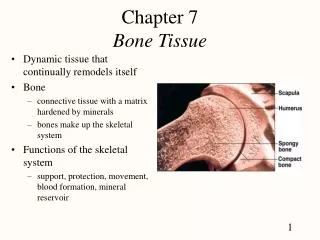
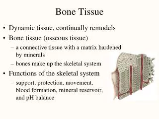
Bone Tissue. BIO 137 Anatomy & Physiology I. The Skeletal System. Organs Bones Cartilages Tendons Ligaments. Functions Support Protection Movement Hematopoiesis Mineral Store Energy Store. Tissues of the Skeletal System.

E N D
Bone Tissue BIO 137 Anatomy & Physiology I
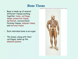
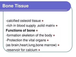
The Skeletal System • Organs • Bones • Cartilages • Tendons • Ligaments • Functions • Support • Protection • Movement • Hematopoiesis • Mineral Store • Energy Store
Tissues of the Skeletal System • Bone is a highly vascularized C.T. with a hard, mineralized extracellular matrix. It is found in the body in two different arrangements: • Compact bone – 80% of bone in skeleton • Spongy bone - 20% of bone in skeleton • is seen as
Compact Bone • Most rigid connective tissue • Continuous, solid extracellular matrix found between cells • Matrix composed of mineral salts and collagen • Hardness due to mineral salts in matrix • Collagen provides strength and resilience
Spongy Bone • Made of Bony plates called trabeculae • Space between trabeculae filled with red bone marrow • Found in flat bones between sheets of compact bone • Found in epiphyses of long bones • Spongy bone reduces a bone’s weight
Long Bone Structure • Epiphysis - Expanded ends of a long bone, articulates with other bones • Articular cartilage (hyaline cartilage) on outer surface • Composed of mostly spongy (cancellous) bone surrounded by thin layers of compact bone • Red bone marrow with Hematopoietic stem cells located here Hyaline cartilage is the articular cartilage of this long bone
Long Bone Structure • Diaphysis– shaft of bone • Central medullary cavity • Hollow tube in diaphysis filled with yellow marrow • Medullary Cavity is lined by endosteum • Osteoclasts located in endosteum • Metaphysis – region between diaphysis and epiphysis • Growth plate here
Lone Bone Structure • The medullary cavity is a space within the diaphysis of long bones that contains fatty yellow bone marrow in adults. • The endosteum is a membrane that lines the medullary cavity . • The endosteum is composed of osteoclasts, osteoblasts, and connective tissue. • Nutrient foramen – canal where vessels enter/exit a bone
Long Bone Structure • The periosteum is a tough sheath of dense, irregular connective tissue on the outside of the bone. • It contains osteoblasts that help the bone grow in thickness, but not in length. • Continuous with tendons and ligaments • It also assists with fracture repair
Chemical Constituents of Bone • Bone is 15% water, 30% organic proteins, 55% mineral salts (hydroxyapatite crystals). • Organic constituents • Collagen fibers provide flexibility and tensile strength. • Inorganic hydroxyapatite crystals (mineral salts) • Calcium Phosphate (Ca3PO4)2 • Calcium Carbonate (CaCO3 – marble) • Other trace elements: magnesium, fluoride, sulfate
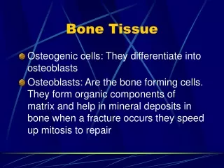
Bone Cells • Osteogenic Stem Cell • Derived from mesenchyme • Forms osteoblasts • Only Mitotic bone cell • Osteoclasts • Bone breakdown (resorption) • Osteoblasts • Bone forming cells, deposit bony matrix around themselves • Osteocytes • matured osteoblasts • Completely surrounded by bony matrix • Carries out daily cellular activities
Histology of Bone Tissue • Compact bone is composed of repeating structural units called Osteons or Haverisan Systems • Osteons have concentric circles of matrix called lamellae surrounding a Central (Haversian) canal • Blood vessels and nerves here • Osteoblast cells mature into Osteocytes after matrix is laid down
Histology of Bone Tissue • Lacunae spaces in osetonshosue osteocytes • Canaliculi– small channels that connect lacunae of osteocytes • Nutrient and waste transport • Gap junctions for communication • Perforating (Volkmann’s) canals – transverse canals that connect vessels of central (Haversian) canals
Blood and Nerve Supply of Bone • Bone is richly supplied with blood; Periosteal arteries and veins supply the periosteum and compact bone. • Nerves accompany the blood vessels • The periosteum is rich in sensory nerves sensitive to tearing or tension
Bone Formation • Ossification or osteogenesis is the process of forming new bone. Bone formation occurs in four situations: • Formation of bone in an embryo • Growth of bones until adulthood • Remodeling of bone • Repair of fractures
Bone Formation • Osteogenesis occurs by two different methods, beginning about the 6th week of embryonic development. • Intra-membranous ossification produces spongy bone. • This bone may subsequently be remodeled to form compact bone. • Endochondralossification is a process whereby cartilage is replaced by bone. • Forms both compact and spongy bone.
Bone Formation • Intra-membranous ossification is the simpler of the two methods. • It is used in forming the flat bones of the skull, mandible, and clavicle. • Bone forms from mesenchymal cells that develop within a membrane – without going through a cartilage stage • Many ossification centers.
Bone Formation • Endochondral ossification is the method used in the formation of most bones, especially long bones. • It involves replacement of hyaline cartilage bone models. • There are one primary and two secondary centers of growth.
Bone Formation • Ossification contributing to bone length is usually complete by 18-21 years of age. • Bones can still continue to thicken and are capable of repair even after the epiphyseal growth plates have closed.
Bone Structure • In adolescents, through the end of active growth, the epiphysis of the long bones contains hyalinecartilage and forms an “epiphysealgrowth plate”. • The growth plate is always actively dividing causing the bone to elongate from each end.
Bone Structure • In adults, the epiphyseal cartilage is no longer present and elongation of bones has stopped. • The epiphyseal growth plate becomes an “epiphyseal line”, as growing cartilage is replaced by calcified bone.
Long Bone Growth at Epiphyseal Plate • 4 zones of cartilage at epiphyseal plate • Zone of Resting cells • Closest to epiphysis, don’t participate in growth, anchors plate to bony tissue of epiphysis • Zone of Mitotic cells • Rows of many cells undergoing mitosis • As ECM forms around cartilage thickens • Zone of Hypertrophic cells • Older cells enlarge and thicken plate, lengthening bone • Calcifying older cells begin to die • Zone of Calcified cartilage • Thin layer of dead cartilage • Osteoblasts lay down bone to replace calcified cartilage
Articular cartilage Epiphysis New chondrocytes are formed EPIPHYSEAL (GROWTH) PLATE: Zone of resting cartilage Zone of proliferating cartilage Old chondrocytes are replaced by bone Zone of hypertrophic cartilage New diaphysis Zone of calcified cartilage Diaphysis (c) Lengthwise growth of bone at epiphyseal plate
Long Bone Growth at Epiphyseal Plate • 2 MAJOR EVENTS 1. New cartilage cells are produced by mitosis at epiphyseal end of plate 2. Replacement of cartilage with bone on diaphyseal end of plate by endochondral ossification • Epiphyseal plate temporarily increases in width • Bones continue to lengthen until ossification centers meet and the epiphyseal plate ossifies under control of sex hormones at end of puberty
Bone Growth in Thickness: Appositional Growth • A bone thickens as Compact bone is deposited below the periosteum • Osteoclasts will resorb (break down) bone on the inside to form the medullary cavity • The diaphysis of a long bone grows in thickness on the outside while the medullary cavity is enlarged on the inside
Bone Growth & Remodeling • Ongoing process that includes bone resorptionby osteoclasts and deposition of boneby osteoblasts • Opposing processes • Total mass of bone in skeleton remains constant • At any one time, 5% of total bone mas is being remodeled
Factors affecting Bone Development, Growth & Repair • Normal bone growth depends on several factors: • Nutrition • Hormonal secretions • Physical exercise
Nutritional Requirements for Bone Development • Vitamin A • Required for normal osteoblast and osteoclast activity during normal development • Vitamin C • Required for collagen synthesis • Vitamin D • Promotes calcium absorption in the small intestines
Factors affecting Bone Development, Growth & Repair • Physical stress from exercise • Stimulates bone growth (deposition) • Lack of exercise makes bones atrophy (weaker) and thinner • Loss of bone mass
Hormones affecting Normal Bone Growth • Thyroid Hormones • Secreted by the thyroid gland • Stimulates osteoblasts • Deficiency: • Stunted growth
Hormones affecting Normal Bone Growth • Sex hormones: Estrogen and Testosterone • Promote formation of bone tissue • Abundant at puberty, stimulate long bone growth • Growth Spurt • Estrogen effect stronger than testosterone • Responsible for closing epiphyseal plates • Widening of female pelvis
Hormone Requirements for Bone Development • Growth hormone – secreted from the anterior pituitary gland • Regulates overall body growth • IGFs released from liver and bone in response to GH • Stimulates division of cartilage cells in epiphyseal plates during growing years and stimulates growth in thickness • Role in metabolism (discussed in Endocrine System)
Growth Hormone Disorders • Pituitary Dwarfism • Pituitary gigantism • Acromegaly
Growth Hormone Deficiency • Pituitary Dwarfism • GH deficiency in children • Short stature from retarded skeletal growth • Normal body proportions
Growth Hormone Excess • Acromegaly • GH excess in adulthood • Bones become thicker & soft tissues proliferate • Disfuguring
Growth Hormone Excess • Pituitary Gigantism • GH excess in childhood before epiphyseal plates have closed • Rapid growth in height
Blood Calcium Homeostasis • Bone is the body’s major calcium reserve, storing 99% of calcium • Inorganic salts in matrix of bone composed mostly of calcium phosphate and calcium carbonate • Calcium required for blood clot formation, nerve impulse conduction and muscle cell contraction • Calcium homeostasis maintained by three hormones: • Parathyroid hormone • Calcitonin • Vitamin D
Blood Calcium Homeostasis • Parathyroid Hormone • Secreted by parathyroid glands in response to low blood calcium • Stimulates osteoclast activity = Bone resorption • Secrete acids and enzymes that dissolve inorganic mineral matrix of bone • Releases calcium and phosphorous into the blood
Parathyroid Hormone • Other PTH targets: • Osteoclasts – Bone resorption • Kidneys • Reabsorb calcium during urine formation • Secrete phosphate during urine formation • Intestines • Increases calcium absorption through activation of Vitamin D regulation • This is necessary for the intestines to absorb Ca++ • Functions to increase the calcium concentration of the plasma (also increases concentration of phosphate)
Blood Calcium Homeostasis • Vitamin D • Necessary for proper absorption of calcium in the small intestines • Lack of vitamin D decreases calcium absorption in small intestines • Decreases calcium content in bone matrix, making bones soft and deformed • Rickets in children • Osteomalacia in adults • Derivative of Cholesterol is converted by sunlight to Vitamin D
Blood Calcium Homeostasis • Calcitonin • Released from thyroid gland in response to high levels of blood calcium • Inhibits osteoclast activity • Stimulates osteoblasts to form bone tissue • Stimulates the kidneys to secrete calcium • Functions to decrease plasma calcium concentration