Download
1 / 35
470 likes | 1.1k Views
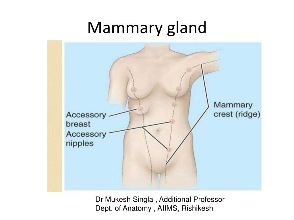
Mammary gland. Dr Mukesh Singla , Additional Professor Dept. of Anatomy , AIIMS, Rishikesh. OBJECTIVES. By the end of the lecture, the student should be able to describe : Shape and position of the female breast. Structure of the mammary gland.

E N D
Mammary gland Dr Mukesh Singla , Additional Professor Dept. of Anatomy , AIIMS, Rishikesh
OBJECTIVES By the end of the lecture, the student should be able to describe: • Shape and position of the female breast. • Structure of the mammary gland. • List blood supply of the female breast. • Lymphatic drainage of the female breast. • Applied anatomy in the female breast
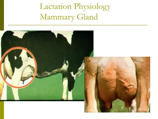
INTRODUCTION • Mammary Glands exist in both sexes. • Rudimentary in males throughout life • Start developing at puberty in females • Most of the development occurs during later months of pregnancy and lactation
Parts, Shape & position of the Gland • It is conical in shape. • It lies in superficial fascia of the front of chest. • It has a base, apex and tail. • Its base extends from 2nd to 6th ribs. • It extends from the sternum to the midaxillary line laterally. • It has no capsule.
SHAPE AND POSITION OF FEMALE BREAST • 2/3 of its base lies on the pectoralis major muscle, while its inferolateral 1/3 lies on: • Serratus anterior & • External oblique muscles. • Its superolateral part sends a process into the axilla called the axillary tail or axillary process.
SHAPE AND POSITION OF FEMALE BREAST • Nipple: • It is a conical eminence that projects forwards from the anterior surface of the breast. • The nipple lies opposite 4th intercostal space. • It carries 15-20 narrow pores of the lactiferous ducts. • Areola : • It is a dark pink brownish circular area of skin that surrounds the nipple. • The subcutaneous tissues of nipple & areola are devoid of fat.
STRUCTURE OF MAMMARY GLAND • It is non capsulated gland. • It has fibrous strands (ligaments of cooper) which connect the skin with deep fascia of pectoralis major. • Retromammary space. What is its Importance?
STRUCTURE OF MAMMARY GLAND • It is formed of 15-20 lobes. • Each lobe is formed of a number of lobules. • It has from 15-20 lactiferous ducts which open by the same number of openings on the summit of the nipple.
ARTERIAL SUPPLY • 1. Perforating branches of internal thoracic (internal mammary) artery. • 2. Mammary branches of lateral thoracic artery. • 3. Mammary branches of Intercostal arteries.
VENOUS SUPPLY • Veins are corresponding to the arteries. • Circular venous plexus are found at the base of nipple. • Finally, veins of this plexus drain into axillary & internal thoracic veins.
AXILLARY LYMPH NODES • They are arranged into 5 groupswhich lie in axillary fat : • Pectoral (Anterior) group : which lies on the pectoralis minoralong lateral thoracic vessels. • Subscapular (Posterior) group : which lies on posterior wall of axilla on lower border of subscapularis along subscapular vessels.
AXILLARY LYMPH NODES • Brachial (Lateral) group : lies on lateralwall of axillaalong 3rd part of axillary vessels. • Central group : lies in axillary fatat the base of axilla. • Apical group : lies at apex of axilla. • Subclavian lymph trunk: • it is formed by union of efferent lymph vessels of apical group. It usually opens in subclavian vein. On the left side it usually opens into thoracic duct.
LYMPHATIC DRAINAGE • Subareolar lymphatic plexus : • Lies beneath the areola. • Deep lymphatic plexus: • Lies on the deep fascia covering pectoralis major. • Both plexuses radiate in many directions and drain into different lymph nodes.
APPLIED ANATOMY- CANCER BREAST • It is a common surgical condition. • 60% of carcinomas of breast occur in the upper lateral quadrant. • 75% of lymph from the breast drains into the axillary lymph nodes. • In case of carcinoma of one breast, the other breast and the opposite axillary lymph nodes are affected because of the anastomosing lymphatics between both breasts.
Applied Anatomy • The lactiferous ducts are radially arranged from the nipple, so incision of the gland should be made in a radial direction to avoid cutting through the ducts. • Infiltration of the ligaments of Cooper by breast cancer leads to its shortening giving peaude’orangeappearance of the breast.
Mammary ridge • Mammary ridge extends from the axilla to the inguinal region. • In human, the ridge disappears EXCEPT for a small part in the pectoral region. • In animals,several mammary glands are formed along this ridge.
Axillary Lymph node • Axillary (ipsilateral): interpectoral (Rotter’s) nodes and lymph nodes along the axillary vein and its tributaries that is divided into the following levels: • Level I (low-axilla): lymph nodes lateral to the lateral border of pectoralis minor muscle. • b. Level II (mid-axilla): lymph nodes between the medial and lateral borders of the pectoralis minor muscle and the interpectoral (Rotter’s) lymph nodes. • c. Level III (apical axilla): lymph nodes medial to the medial margin of the pectoralis minor muscle and inferior to the clavicle. These are also known as apical or infraclavicular nodes.
Sentinel node biopsy and axillary dissection • Sentinel node biopsy is the most common way to check the axillary lymph nodes for cancer. • Before or during the procedure, a radioactive substance (called a tracer) and/or a blue dye is injected into the breast. These substances help the surgeon find the nodes to remove. • The first lymph node(s) to absorb the tracer or dye is called the sentinel node(s). This is also the first lymph node(s) where breast cancer is likely to spread.
Sentinel node biopsy and axillary dissection • The surgeon removes the sentinel node(s) to get it checked if the node(s) contain cancer cells. • If cancer is not found, it’s likely the other nodes do not contain cancer. So, no more surgery is needed. • If the node(s) do contain cancer, more lymph nodes may be removed, which is called axillary dissection.
Lymphedema • When lymph nodes are removed, some of the lymph vessels can become blocked and cause lymphedema. Lymphedema is a build-up of lymphatic fluid. It causes swelling in the arm or other areas such as the hand, fingers, breast, chest or back.
Lymphedema • Lymphedema isn’t common when only a few lymph nodes are removed. The cases that do occur are less severe than when more nodes are removed. • Today, sentinel node biopsy is the preferred way to remove lymph nodes (only a few nodes are removed). So, most people don’t get lymphedema.
Clinical anatomy • Skin incisions over breast • Retraction of skin and nipple • Congenital anomalies- ‘’thelia’’ and ‘’mastia’’ • Lymphadenopathy • Krukenberg’s tumour • Breast examination
1- • A 40 years female presents with pain along the medial side of arm. On examination- she has palpable lump in upper outer quadrant of breast and enlarged axillary lymph nodes. This referred pain is due to enlarged lymph nodes compressing- • A- long thoracic nerve • B- Intercostobrachial nerve • C- lateral pectoral nerve • D- medial cutaneous nerve of arm
2 • Which of the following is correct about lymphatic drainage of mammary gland? • A- pectoral group of lymph nodes lie along lower border of pectoralis major • B-medial group of axillary lymph nodes drain inner quadrants • C- 75% of lymphatics from mammary glands drained by Internal mammary nodes • D- lymphatics from breast can transmit to ovarian surface