Download
1 / 16
230 likes | 884 Views
Thyroid gland. Introduction- Dimension- Each lobe 5x2.5x2.5cm Isthmus- 1.2x1.2 cm 12g average weight . Thyroid gland. Situation and extent- C5,C6,C7&T1; embracing the upper part of the trachea Each lobe extends from the middle of the thyroid cartilage up to 5 th or 6 th tracheal ring

E N D
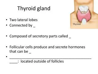
Thyroid gland Introduction- Dimension- Each lobe 5x2.5x2.5cm Isthmus- 1.2x1.2 cm 12g average weight
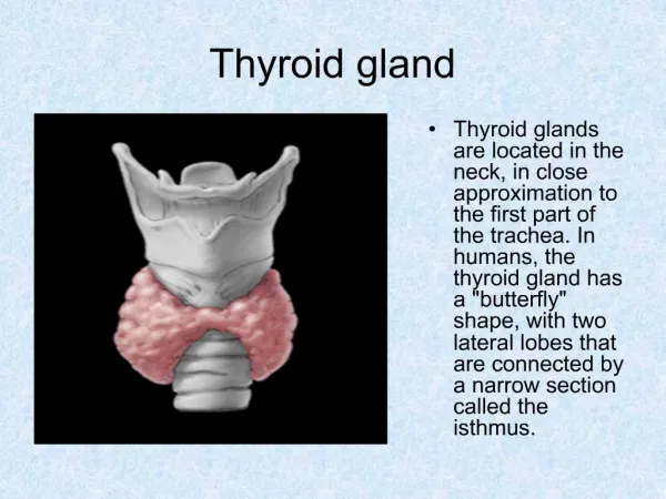
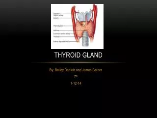
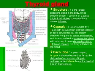
Thyroid gland Situation and extent- • C5,C6,C7&T1; embracing the upper part of the trachea • Each lobe extends from the middle of the thyroid cartilage up to 5th or 6th tracheal ring • Isthmus extends from the 2nd to 4th tracheal ring
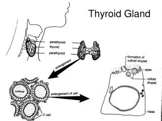
Thyroid gland Capsule of the thyroid gland- • True capsule- is peripheral condensation of the connective tissue of the gland • False capsule- pretracheal fascia of the deep cervical fascia. Thin along the posterior border of the lobes but thick on the inner surface of the gland- suspensory lig ( of Berry) which connects the lobe to the cricoid cartilage. Dense capillary plexus is present deep to the true capsule
Thyroid gland Relations- • Apex • Base • 3 surfaces, lateral, medial and posterolateral • 2 borders- ant & post
Thyroid gland Apex- directed upwards & slightly laterally, superiorly limited by the attachment of the sternothyroid to the oblique line of the thyroid cartilage Base- 4th to 5th tracheal ring Lateral surface- sternohyoid, superior belly of omohyoid, sternothyroid and ant border of the sternocleidomastiod muscle. Medial surface- two tubes tracheal and oesophagus, two muscles inferior constrictor and cricothyroid & two nerves external laryngeal and recurrent laryngeal Posterolateral- carotid sheath Anterior border- thin &related to ant branch of the superior thyroid artery Posterior border- thick & rounded, related inferior thyroid artery, anastomosis betwn superior and inferior thyroid arteries, parathyroid glands and thoracic duct in the left side
Thyroid gland Isthmus- • 2 surfaces ant & post • 2 borders superior and inferior Ant surface- rt & lt sternohyoid & sternothyroid muscles. Ant jugular veins , fascia and skin. Post surface- 2nd to 4th tracheal rings Superior – anastomosis betwn rt and lt superior thyroid arteries Inferior border- inferior thyroid veins
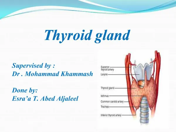
Thyroid gland Arterial supply- • Superior thyroid artery • Inferior thyroid artery • Thyroidea ima artery • Accessory thyroid artery
Thyroid gland Venous drainage- • Superior thyroid vein • Middle thyroid vein • Inferior thyroid vein • Fourth thyroid vein( vein of Kocher)
Thyroid gland Lymphatic drainage- Upper part of the gland- deep cervical lymph nodes either directly or through the prelaryngeal nodes Lower part of the gland to the lower deep cervical lymph nodes directly and also through the pretracheal and paratracheal nodes Nerve supply- Middle cervical ganglion and partly from the superior and inferior cervical ganglion