Download
1 / 51
610 likes | 705 Views
MAMMARY GLAND. This resource is licensed under the Creative Commons Attribution Non-Commercial & No Derivative Works License. Objectives. For the Gross anatomy and Histology of the mammary gland. Students should be able to: Appreciate the differences in duct numbers per teat.

E N D
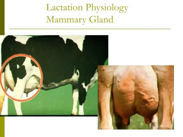
MAMMARY GLAND This resource is licensed under the Creative Commons Attribution Non-Commercial & No Derivative Works License
Objectives For the Gross anatomy and Histology of the mammary gland. Students should be able to: Appreciate the differences in duct numbers per teat. Identify the mammary glands and their main features in all domestic species. Identify their blood vessels, nerves and lymph ducts; appreciate internal structure features. Identify the features of the microscopic structure of active and inactive mammary glands.
SLIDE 98 Mammary gland - inactive At low magnification identify : 1. area of tubules. 2. interlobular connective tissue. 3. lactiferous ducts. 4. teat. 5. surface of skin. 2.5 mm
SLIDE 98 Mammary gland - inactive 5 1 2 At low magnification identify : 1. area of tubules. 2. interlobular connective tissue. 3. lactiferous ducts. 4. teat. 5. surface of skin. 2 3 Connective tissue is stains blue/green in this triple stained section. 1 4 2.5 mm
SLIDE 98 Mammary gland - inactive Area of teat identify : lactiferous ducts. lactiferous sinuses. teat sinus. 1.0 mm
SLIDE 98 Mammary gland - inactive lactiferous sinus ‘star’ shaped appearance Area of teat identify : lactiferous ducts. lactiferous sinuses. teat sinus. connective tissue teat sinus lactiferous ducts 1.0 mm
SLIDE 98 Mammary gland - inactive Opening of teat sinus, identify : teat sinus. glands opening into sinus. keratinised, stratified squamous epithelium. 250 µm
SLIDE 98 Mammary gland - inactive keratinised stratified squamous epithelium fold in section fold Opening of teat sinus, identify : teat sinus. glands opening into sinus. keratinised, stratified squamous epithelium. glands teat canal teat sinus 250 µm
SLIDE 98 Mammary gland - inactive Identify : lobules interlobular duct system interlobular connective tissue 0.5 mm
SLIDE 98 Mammary gland - inactive interlobular ducts opening to lactiferous ducts Ct : interlobular connective tissue L L : lobules Ct Identify : lobules interlobular duct system interlobular connective tissue L L lactiferous duct Ct 0.5 mm
SLIDE 98 Mammary gland - inactive At a slightly higher magnification the inactive tubules forming the lobules of the gland can be identified. Large areas of interlobular connective tissue, stained blue/green extend between these lobules. Within this connective tissue run the interlobular ducts. 250 µm
SLIDE 98 Mammary gland - inactive connective tissue At a slightly higher magnification the inactive tubules forming the lobules of the gland can be identified. Large areas of interlobular connective tissue, stained blue/green extend between these lobules. Within this connective tissue run the interlobular ducts. interlobular duct lobule 250 µm
SLIDE 98 Mammary gland - inactive The low columnar cells lining the walls of the tubules are mildly secretory - note the eosinophilic content of some of the tubules. 50 µm
SLIDE 98 Mammary gland - inactive S : secretions in tubules S S The low columnar cells lining the walls of the tubules are mildly secretory - note the eosinophilic content of some of the tubules. S 50 µm
SLIDE 98 Mammary gland - inactive How is the inactive mammary gland classified? 50 µm
How is the inactive mammary gland classified? Compound tubular gland. SLIDE 98 Mammary gland - inactive tubules intralobular connective tissue How is the inactive mammary gland classified? interlobular connective tissue blood vessel 50 µm
SLIDE 98 Mammary gland - inactive What are the elongated cells with nuclei lying at the base of the tubule epithelium? 25 µm
SLIDE 98 Mammary gland - inactive Arrowed : myoepithelial cells T : tubules T Ct : connective tissue (intralobular) What are the elongated cells with nuclei lying at the base of the tubule epithelium? Myoepithelial cells. Ct S : secretion T S 25 µm
SLIDE 98 Mammary gland - inactive Towards the teat the lactiferous ducts open into the lactiferous sinuses to teat sinus to teat canal. The double epithelial layers become progressively taller, from cuboidal to columnar. 0.5 mm
SLIDE 98 Mammary gland - inactive What is a distinguishing feature of the lactiferous ducts? 50 µm
What is a distinguishing feature of the lactiferous ducts? Double layered cuboidal to columnar epithelium. SLIDE 98 Mammary gland - inactive double layered epithelium What is a distinguishing feature of the lactiferous ducts? lumen of duct connective tissue 50 µm
Demonstration slide Mammary gland Transverse section through teat canal. Note : keratinized stratified squamous epithelium lining the canal, bands of smooth muscle and blood vessels.
Demonstration slide Mammary gland blood vessels epithelium keratinised stratified squamous Transverse section through teat canal. Note : keratinized stratified squamous epithelium lining the canal, bands of smooth muscle and blood vessels. smooth muscle
SLIDE 99 Mammary gland - active If you look at slide 99 in the class; start by examining the red/pink stained section. 0.5 mm
SLIDE 99 Mammary gland - active Note each lobule is greatly expanded; consisting mostly of alveoli. The interlobular connective tissue is compressed to fibrous tracts or seams. 250 µm
SLIDE 99 Mammary gland - active Ct : connective tissue A : alveoli Ct Ct A Note each lobule is greatly expanded; consisting mostly of alveoli. The interlobular connective tissue is compressed to fibrous tracts or seams. adipocytes A A Ct interlobular duct 250 µm
SLIDE 99 Mammary gland - active Identify : 1. lobule. 4. alveoli. 2. intralobular ducts. 5. interlobular ducts. 3 interlobular connective tissue. 250 µm
SLIDE 99 Mammary gland - active 4 4 1 2 3 Identify : 1. lobule. 4. alveoli. 2. intralobular ducts. 5. interlobular ducts. 3 interlobular connective tissue. 4 3 3 5 250 µm
SLIDE 99 Mammary gland - active In the compressed seams of interlobular connective tissue, Identify : blood vessels. unilocular adipocytes. 100 µm
SLIDE 99 Mammary gland - active arteriole unilocular adipocytes alveoli In the compressed seams of interlobular connective tissue, Identify : blood vessels. unilocular adipocytes. connective tissue (interlobular) 100 µm
SLIDE 99 Mammary gland - active Most prominent in the interlobular connective tissue are the large interlobular and lactiferous ducts. 50 µm
SLIDE 99 Mammary gland - active alveolar epithelium alveolus blood vessels Most prominent in the interlobular connective tissue are the large interlobular and lactiferous ducts. interlobular connective tissue interlobular duct note cuboidal/columnar epithelium 50 µm
SLIDE 99 Mammary gland - active Within the lobules identify intralobular ducts. 50 µm
Within the lobules identify intralobular ducts. Note the more regular epithelium of the duct. SLIDE 99 Mammary gland - active alveoli Within the lobules identify intralobular ducts. intralobular duct 50 µm
SLIDE 99 Mammary gland - active How is the active mammary gland classified? 50 µm
How is the active mammary gland classified? Compound tubulo-alveolar gland. SLIDE 99 Mammary gland - active alveoli How is the active mammary gland classified? connective tissue seams 50 µm
SLIDE 99 Mammary gland - active Note the appearance of the alveoli : folded somewhat ‘ragged’ epithelium. content of fat droplets. eosinophilic protein containing secretions. 50 µm
SLIDE 99 Mammary gland - active E E : eosinophilic secretions E Note the appearance of the alveoli : folded somewhat ‘ragged’ epithelium. content of fat droplets. eosinophilic protein containing secretions. adipocytes folded epithelium 50 µm
SLIDE 99 Mammary gland - active Compare the staining of the large unilocular adipocytes in this section with their appearance in the lipid stained section which follows. 50 µm
SLIDE 99 Mammary gland - active unilocular adipocytes in seam of interlobular connective tissue Compare the staining of the large unilocular adipocytes in this section with their appearance in the lipid stained section which follows. When inactive; the alveolar cells are phagocytosed by invading macrophages. alveolar epithelium 50 µm
SLIDE 99 Mammary gland – active stained for lipid As seen before in histological sections; the presence of lipid can be demonstrated by the use of osmium containing solutions. In sections stained with H&E the unfixed lipid is lost during sample processing. 1.0 mm
SLIDE 99 Mammary gland – active stained for lipid As seen before in histological sections; the presence of lipid can be demonstrated by the use of osmium containing solutions. In sections stained with H&E the unfixed lipid is lost during sample processing. All handling and use of osmium containing solutions must be carried out in a fume hood. 1.0 mm
SLIDE 99 Mammary gland – active stained for lipid Compare the small globules of fat in the alveoli with the larger, darker globules of the adipose cells in the interlobular connective tissue. 250 µm
SLIDE 99 Mammary gland – active stained for lipid interlobular connective tissue A : alveolae Ua : unilocular adipocytes A Compare the small globules of fat in the alveoli with the larger, darker globules of the adipose cells in the interlobular connective tissue. A Ua Ua 250 µm
SLIDE 99 Mammary gland – active stained for lipid When viewing this section examine under higher magnification, look at a brown/black area rather than intensely black over-stained areas. 100 µm
SLIDE 99 Mammary gland – active stained for lipid F : fat globules interlobular connective tissue F epithelium F When viewing this section examine under higher magnification, look at a brown/black area rather than intensely black over-stained areas. alveoli adipocytes 100 µm
SLIDE 99 Mammary gland – active stained for lipid Then at still higher magnification look for fat globules in the alveoli and small globules actually in the apical parts of the alveolar epithelial cells. 50 µm
SLIDE 99 Mammary gland – active stained for lipid cell nucleus Then at still higher magnification look for fat globules in the alveoli and small globules actually in the apical parts of the alveolar epithelial cells. droplets in apical area of cell free lipid droplets alveolar lumen 50 µm
SLIDE 99 Mammary gland – active stained for lipid Why would a stain dissolved in water stain fat sections. 50 µm
Why would a stain dissolved in water stain fat sections. The stain is more readily soluble in the fat than in water. SLIDE 99 Mammary gland – active stained for lipid lipid droplets in cell Why would a stain dissolved in water stain fat sections. alveolar lumen 50 µm