Download
1 / 63
790 likes | 2.86k Views
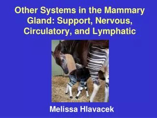
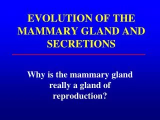
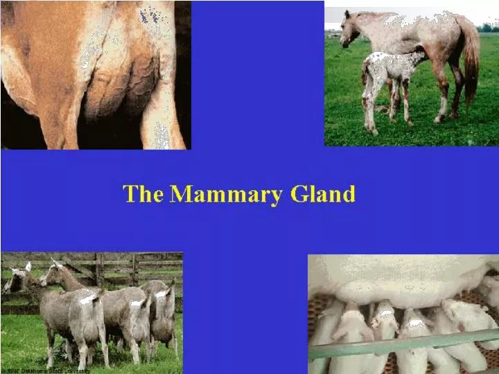
The Mammary Gland. The milk secreting organ Modified sweat gland Exocrine gland. MAMMARY GLANDS. thoracic. inguinal. abdominal. MAMMARY GLANDS. 4 abdominal; 15 openings / teat. MAMMARY GLANDS. 2 thoracic, 6 abdominal, ; 3-7 openings / teat. MAMMARY GLANDS.

E N D
The Mammary Gland • The milk secreting organ • Modified sweat gland • Exocrine gland
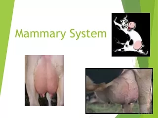
MAMMARY GLANDS thoracic inguinal abdominal
MAMMARY GLANDS • 4 abdominal; 15 openings / teat
MAMMARY GLANDS • 2 thoracic, 6 abdominal, ; 3-7 openings / teat
MAMMARY GLANDS • 2 thor., 6 abdom. 2 ing., ; 8-10 openings/ teat
MAMMARY GLANDS • 4 thor., 2 abdom. 4 ing., ; 1 opening/ teat
MAMMARY GLANDS • 2 inguinal ; 1 opening/ teat
MAMMARY GLANDS • 2 inguinal ; 2 openings/ teat
MAMMARY GLANDS • 4 thor., 6 abdom. 2 ing., ; 2 openings/ teat
MAMMARY GLANDS • 4 inguinal ; 1 opening/ teat
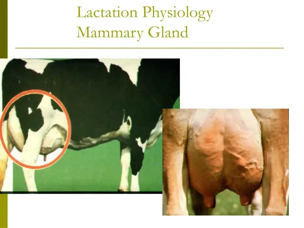
Anatomy of the Mammary Gland • Mammary gland - milk secreting structure including teats, duct system, lobes, lobules, and secretory tissue • Modified sweat gland • Exocrine gland • Cow • Large & in inguinal region • 4 teats/quarters = 4 separate glands • No mixing of ducts across quarters
Anatomy of the Mammary Gland • Rear quartes produce approx. 60% of the milk and the fore quarters produce the remaining 40% • The size and shape of udders vary with the 1) producing ability, 2) age, and 3) genetic of the cow
Anatomy of the Mammary Gland • Cow • Front & rear quarters separated by fine membrane • Left and right separated by median suspensory ligament • Supernumerary teats (some with duct and secretory system)
How much support is enough? • High producing Holstein cow • Empty Udder = 25 kg. • Milk = 30 kg • 25 + 30 = 55 kg !!!
Udder Support in Cow • Skin • Fine connective tissue below skin • Connective tissue attaches front quarters to abdominal wall • Lateral suspensory ligaments (LSL) • Median suspensory ligament (MSL) • The subpelvic tendon
Udder Support in Cow • LSL • Sling around udder • 2 layers • Inelastic, more fibrous than MSL
Lateral Suspensory Ligaments • Like a “hammock” around the udder • From the pelvis to the median suspensory ligament • Mostly fibrous tissue • Collagen • Attaches to the alveolar tissue • Provides internal framework
Udder Support in Cow • MSL • Primary support • Relatively elastic • 2 layers • Broken MSL – pendulous udder
Median Suspensory Ligament • Primary support of the udder • Two adjacent heavy sheets of tissue • Mostly elastic, some fibrous tissue • Attaches to the abdominal wall • Divides the udder into halves • Glands on each half are divided by sheetsof tissue © Biology of Lactation, Schmidt
Udder Support in Cow • Lamella septa • Connective tissue • Runs between LSL & MSL • Divides parenchyma into lobes and lobules
Duct System • Teat meatus, the small canal located in the end of each teat is .5 to 1 cm long and is the only sphincter in each gland • Seven or eight loose folds of membrane known as furstenburg rosette are located above the teat meatus • The teat cistern, the cavity within the teat hold 30 to 90 ml of milk.
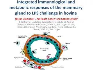
The Secretory Tissue • A Lobe: group of lobules • A Lobule: group of alveoli • Alveoli: cluster of alveolus • Alveolus: a single layer of epithelial cells surrounding a central lumen
Blood Supply to Mammary Gland • 400 kg blood to produce 1 kg of milk • 2 major arteries • Front ½ of udder • Rear ½ of udder • 4 major veins • 2 follow same path as arteries • 2 mammary veins
Nervous System • Sensory (afferent) nerves in skin and teats • Positive stimulation of teats and surrounding area initiates milk let-down reflex via oxytocin © Biology of Lactation, Schmidt
Nervous System • Sympathetic (efferent) (involuntary) nerves associated with arteries in the gland • Control blood flow to the gland • Innervation of sphincters muscles in teats • Stress causes vasoconstriction decreasing milk secretion and let-down • No parasympathetic innervation • No nerves to myoepithelial cells or alveolar cells
Lymph System of Mammary Gland • What is lymph & what does it do? • Supramammary lymph nodes • Lymph vessels • Factors that influence edema • Age • Diet (especially NaCl) • Exercise • Genetics
Mammary Gland Development • Five phases of mammary development • Prenatal (teats & cisterns dev.) • Prepubertal (limited growth) • Postpubertal • Pregnancy (most growth) • Early lactation
Mammary Gland Development • Major development occurs at puberty and during gestation • Hormones • Estrogen (growth of duct system) • Progesterone (development of alveolar tissue in combination with other hormones) • GH (growth of duct system) • Prolactin (initiation and continuity of lactation)