Download
1 / 28
290 likes | 624 Views
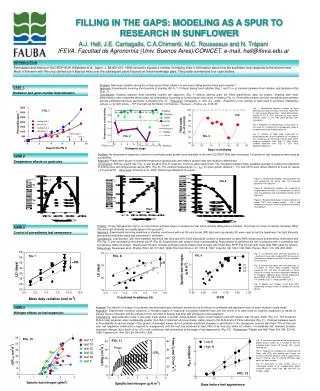
Fig. 17-5. Second mRNA base. First mRNA base (5 end of codon). Third mRNA base (3 end of codon). the mechanism of translation. Amino acids. Polypeptide. tRNA with amino acid attached. Ribosome. Trp. Phe. Gly. tRNA. Anticodon. Codons. 5 . 3 . mRNA. 3 . Amino acid

E N D
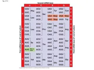
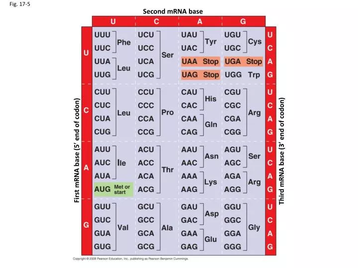
Fig. 17-5 Second mRNA base First mRNA base (5 end of codon) Third mRNA base (3 end of codon)
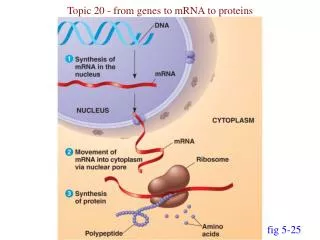
the mechanism of translation Amino acids Polypeptide tRNA with amino acid attached Ribosome Trp Phe Gly tRNA Anticodon Codons 5 3 mRNA
3 Amino acid attachment site 5 Fig. 17-14 Hydrogen bonds Anticodon (a) Two-dimensional structure Amino acid attachment site 5 3 Hydrogen bonds 3 5 Anticodon Anticodon (c) Symbol used in this book (b) Three-dimensional structure
Aminoacyl-tRNA synthetase (enzyme) Amino acid • Attaching amino acids to tRNAs: • Amino-acyl tRNA synthases • 20 different synthases • Require ATP • Each must be specific to the right amino acid and tRNA(s) P P P Adenosine ATP P Adenosine tRNA P P i Aminoacyl-tRNA synthetase P i P i tRNA P Adenosine AMP Computer model Aminoacyl-tRNA (“charged tRNA”)
Aminoacyl-tRNA synthase (ATSGLN) tRNAGLN Adenylated Glutamine
P site (Peptidyl-tRNA binding site) A site (Aminoacyl- tRNA binding site) E site (Exit site) E P A Large subunit mRNA binding site Small subunit Fig. 17-16b (b) Schematic model showing binding sites Growing polypeptide Amino end Next amino acid to be added to polypeptide chain E tRNA mRNA 3 Codons 5 (c) Schematic model with mRNA and tRNA
The Ribosome LSU SSU
Amino end of polypeptide E 3 mRNA P site A site 5 Fig. 17-18-1
Amino end of polypeptide E 3 mRNA P site A site 5 GTP Fig. 17-18-2 GDP E A P
Amino end of polypeptide E 3 mRNA P site A site 5 GTP Fig. 17-18-3 GDP E A P E P A
Peptide bond formation - Transfer of growing chain from tRNA in P site to tRNA in A site
Amino end of polypeptide E 3 mRNA P site A site 5 GTP Fig. 17-18-3 GDP E A P E P A
Amino end of polypeptide E 3 mRNA P site A site Ribosome ready for next aminoacyl tRNA 5 GTP Fig. 17-18-4 GDP E E P A A P GDP GTP E P A
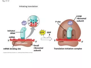
Initiating translation Large ribosomal subunit 3 U 5 C A P site Fig. 17-17 Met Met 5 3 A G U Initiator tRNA GDP GTP E A mRNA 5 5 3 3 Start codon Small ribosomal subunit Translation initiation complex mRNA binding site
Terminating translation Fig. 17-19-1 Release factor 3 5 Stop codon (UAG, UAA, or UGA)
Terminating translation Fig. 17-19-2 Release factor Free polypeptide 3 3 2 GTP 5 5 Stop codon (UAG, UAA, or UGA) 2 GDP
Terminating translation Fig. 17-19-3 Release factor Free polypeptide 5 3 3 3 2 GTP 5 5 Stop codon (UAG, UAA, or UGA) 2 GDP
Completed polypeptide Growing polypeptides Incoming ribosomal subunits Polyribosome Fig. 17-20 Start of mRNA (5 end) End of mRNA (3 end) (a) Ribosomes mRNA (b) 0.1 µm
Second mRNA base The genetic code: -read in triplet codons -once a start codon is specified, codons are read in order (5’ to 3) until a “stop” codon is read -redundant -unambiguous -universal* First mRNA base (5 end of codon) Third mRNA base (3 end of codon)
DNA sequence tRNAGLN RNA sequence Aminoacyl-tRNAsynthase (ATSGLN) Amino acid sequence Adenylated Glutamine Protein structure and function
Altered DNA sequence tRNAGLN Altered RNA sequence Aminoacyl-tRNAsynthase (ATSGLN) Altered (?) Amino acid sequence Adenylated Glutamine Altered (?) Protein structure and function
Fig. 5-17 Nonpolar The 20 amino acids Glycine (Gly or G) Valine (Val or V) Leucine (Leu or L) Isoleucine (Ile or I) Alanine (Ala or A) Trypotphan (Trp or W) Methionine (Met or M) Phenylalanine (Phe or F) Proline (Pro or P) Polar Glutamine (Gln or Q) Serine (Ser or S) Threonine (Thr or T) Cysteine (Cys or C) Tyrosine (Tyr or Y) Asparagine (Asn or N) Electrically charged Acidic Basic Glutamic acid (Glu or E) Histidine (His or H) Aspartic acid (Asp or D) Lysine (Lys or K) Arginine (Arg or R)
Fig. 17-23a Wild type 3 5 DNA template strand 5 3 5 3 mRNA Protein Stop Amino end Carboxyl end A instead of G 5 3 5 3 U instead of C 5 3 Stop Silent (no effect on amino acid sequence)
Fig. 17-23a Wild type 3 5 DNA template strand 5 3 5 3 mRNA Protein Stop Amino end Carboxyl end A instead of G 5 3 5 3 U instead of C 5 3 Stop Silent (no effect on amino acid sequence)
Fig. 17-23b Wild type 3 5 DNA template strand 5 3 5 3 mRNA Protein Stop Amino end Carboxyl end T instead of C 5 3 5 3 A instead of G 3 5 Stop Missense
Fig. 17-22 Wild-type hemoglobin DNA Mutant hemoglobin DNA C T T C 3 3 A T 5 5 T 5 A G G A A 3 5 3 mRNA mRNA G A A 5 G U A 3 5 3 Normal hemoglobin Sickle-cell hemoglobin Val Glu
Fig. 17-23c Wild type DNA template strand 3 5 5 3 mRNA 5 3 Protein Stop Amino end Carboxyl end A instead of T 3 5 3 5 U instead of A 5 3 Stop Nonsense
Fig. 17-23e Wild type 5 DNA template strand 3 5 3 3 mRNA 5 Protein Stop Amino end Carboxyl end missing 5 3 3 5 missing 5 3 Frameshift causing extensive missense (1 base-pair deletion)