Download
1 / 34
520 likes | 793 Views
Smooth muscle. Smooth Muscle. A. fusiform fibers B. centrally located nucleus Few mitochondria . Z-lines are replaced by dense bodies. Sustained contraction. Single unit and multiunit smooth muscles. Smooth muscles are nonstriated and involuntary muscles.

E N D
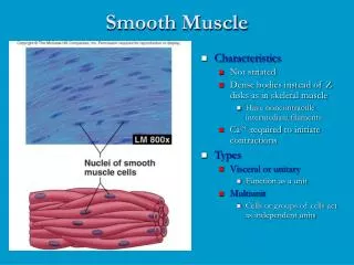
Smooth Muscle • A. fusiform fibers • B. centrally located nucleus • Few mitochondria . • Z-lines are replaced by dense bodies. • Sustained contraction. • Single unit and multiunit smooth muscles.
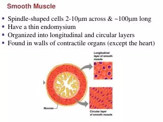
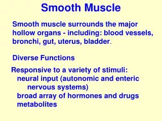
Smooth muscles are nonstriated and involuntary muscles. • Present in almost all the organs in the form of sheets and bundles • Form the major contractile tissues of various organs
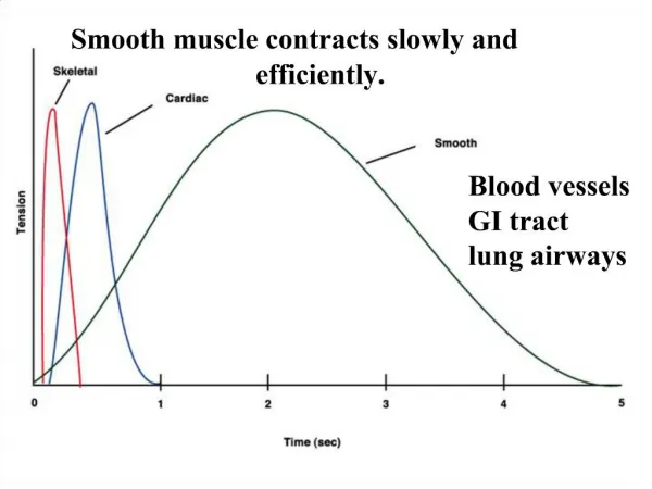
Located in the blood vessels, the respiratory tract, the iris of the eye, the gastro-intestinal tract • The contractions are slow and uniform • Functions to alter the activity of various body parts to meet the needs of the body at that time • Is fatigue resistant • Activation is involuntary Sport Books Publisher
FUNCTION OF SMOOTH MUSCLE • Cardiovascular system • Respiratory system • Digestive system • Renal system
FUNCTIONAL ORGANISATION • Circular • Circular & longitudianal • Circular longitudinal & oblique
Myofilaments of the cells are not parallel as they are in skeletal and cardiac muscle • Symmetrical patterns of thick and thin filaments not evident • No troponincomplex • Contraction is longer and variable and produce less tension. • Types • Single unit and multi unit smooth muscles.
Contract rhythmically as a unit (as one) • Are electrically coupled to one another via gap junctions • Often exhibit spontaneous action potentials • Are arranged in opposing sheets and exhibit stress-relaxation response • Contractile response independent of its innervations • Wall of hollow viscera e.g. GIT, uterus and urinary bladder
MULTI UNIT • Individual units. • Non syncytial • Has its own nerve supply • Contract in response to stimulus • Irregular tetanic contraction. • Location: iris ,ciliary muscles of eye,pilomotor muscle of skin. • Does not respond to stretch.
Spike or AP with prolonged plateau. • Junctional potential • Pacemaker potential
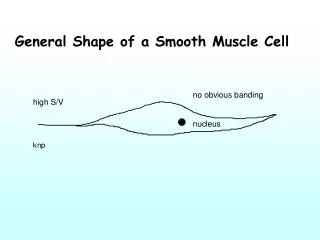
STRUCTURE • Cells are not striated • Fibers smaller than those in skeletal muscle • Spindle-shaped; central nucleus • More actin than myosin • No sarcomeres • Not arranged as symmetrically as in skeletal muscle, thus no striations. • Caveolae: indentations in sarcolemma; • May act like T tubules • Dense bodies instead of Z disks • Have noncontractile intermediate filaments
Smooth Muscle Fibers • Compared to skeletal muscle fibers • shorter • single nucleus • elongated with tapering ends • myofilaments randomly organized • no striations • lack transverse tubules • sarcoplasmicreticulum not well developed
Smooth muscle contraction • Smooth muscle contraction is not controlled by the binding of Ca2+ to the troponin complex as it is in cardiac and skeletal muscles • Calmodulin= intracellular second messenger that binds Ca2+
Smooth Muscle Contraction: Mechanism Figure 12-28: Smooth muscle contraction
Smooth Muscle Relaxation: Mechanism Figure 12-29: Relaxation in smooth muscle
Latch –Bridge mechanism • Attached cross-bridges following dephosphorylation. • Rate of cross-bridge cycle is decreased due to slower rate detachment of cross-bridges. • ATP utilization is less. • Sustained contraction with less energy.
Ephaptic Conduction • Smooth muscle cells are linked electrically via gap junctions so they can stimulate each other causing a wave like action potential • This is also referred to as a FUNCTIONAL SYNCYTIUM
Mechanical properties Tonus Plasticity Slow and prolonged contractile response Length –tension relationship Force-velocity relationship
Muscle tone State of spontaneous irregular contraction. Basic slow wave rhythm. Length-tension relationship Variable
Plasticity • Stress relaxation property of smooth muscle • allows muscle to adjust to stretching without putting undo pressure on contents of organ • a constant state of partial contraction
Response to Stretch • Smooth muscle exhibits a phenomenon called stress-relaxation response in which: • Smooth muscle responds to stretch only briefly, and then adapts to its new length • The new length, however, retains its ability to contract • This enables organs such as the stomach and bladder to temporarily store contents
Force-velocity relationship Low myosin ATPase activity, less no of cross-bridges and slower rate of cross- bridge cycling. Slower rate of cross-bridge cycling.
Neural and Hormonal control ANS influences the pacemaker activity of smooth muscle. Parasympathetic control. Excitatory Acetyl-choline –stimulatory MP decreases-increase the frequency of Aps Increased tonic contractions
Sympathetic • Inhibitory • Releases of catecholamines • Increase MP • Relaxation of muscle • NE-relaxation • Atropine –relaxation • NO, Angiotensin II ,vasopressin stimulate contraction of smooth muscle. • Cold ,stretch stimulate contraction • Hypoxia ,hypercapnia- relaxation of smooth muscle.
Cardiac Muscle and Heart Function • Fibers are branched; connect to one another at intercalated discs. The discs contain several gap junctions • SR is less abundant than in skeletal muscle, but greater in density than smooth muscle • Sarcolemma has specialized ion channels • Fibers are not anchored at ends; allows for greater sarcomere shortening and lengthening
There are different types of cardiac muscle cells ranging from the pacemaker cells in the sinoatrial node to the atrial and ventricular cells that produce the contraction of the heart chambers Pacemaker Cells Have unstable resting membrane potential (pacemaker potential) that slowly drifts upwards until it reaches a threshold and activates and action potential
Phase 0: • Activation of fast Na+ channel-- initial depolarization; • Phase 1: • Partial repolarization; K+ efflux • Phase 2: • Ca2+ entry with continued K+ efflux = "plateau phase". Initial Ca2+ influx through slow L- type Ca2+ • Phase 3 : • This phase is dominated by K+ efflux, i.e. repolarization. • Phase 4 : • This phase is between action potentials)
3 types of muscle Smoot Cardiac Skeletal