Download
1 / 31
380 likes | 1.16k Views
Smooth muscle Contraction. Spindle Shaped Central nuclei Lack Striations, transverse tubules, and lack well developed sacroplasmic reticulum Actin and myosin thin and randomly distributed Multi-unit-Separate units Muscle of iris and blood vessels Visceral-Sheets of spindle cells

E N D
Smooth muscle Contraction • Spindle Shaped • Central nuclei • Lack Striations, transverse tubules, and lack well developed sacroplasmic reticulum • Actin and myosin thin and randomly distributed • Multi-unit-Separate units • Muscle of iris and blood vessels • Visceral-Sheets of spindle cells • Respond as a single unit • Rhythmicity- spontaneous • peristalsis
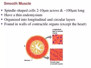
Internal organs- • outer longitudinal • Inner circular Contraction Impulses travel across the membrane- Calcium diffuses into the cell from the extracellular fluid. Calcium binds to a protein calmodulin-activating contraction. Nt- Ach and Norepi, stimulates/ inhibits contractions altering degrees of contraction. Smooth- slow and longer contraction
Terms • Origin • Insertion • Prime mover-Agonist • Assist a prime mover-Synergists • Antagonist- movement in the opposite direction or resist a prime mover,
Skeletal Muscle Actions • origin – immovable end • insertion – movable end • prime mover (agonist) – primarily responsible for movement • synergists – assist prime mover • antagonist – resist prime mover’s action and cause movement in the opposite direction
Muscular Tissue Three Types of Muscle Tissues • Cardiac Muscle • wall of heart • not under conscious control • striated • Skeletal Muscle • usually attached to bones • under conscious control • striated • Smooth Muscle • walls of most viscera, blood vessels, skin • not under conscious control • not striated
Smooth Muscle Fibers • Compared to skeletal muscle fibers • shorter • single nucleus • elongated with tapering ends • myofilaments randomly organized • no striations • lack transverse tubules • sarcoplasmic reticula not well developed
Cardiac Muscle • only in the heart • muscle fibers joined together by intercalated discs • fibers branch • network of fibers contracts as a unit • self-exciting and rhythmic • longer refractory period than skeletal muscle
Orbicularis oculi-blinking/closes eyelids Orbicularis oris-closes lips/protrudes lips-kissing Buccinator-compresses cheeks as in blowing air Zygomaticus-raises corner of mouth/smiling Platysma-pouting/draws mouth downward & elevates skin Frontalis-elevates eyebrows & creases skin Muscles of Facial Expression-CNVII
Masseter-elevates mandible Temporalis-elevates mandible Pterygoid Medial-elevates and moves it from side to side Lateral-depresses and protracts Muscles of Mastication-CNV3
Muscles of Head and Vertebra • Paired muscles- flex, extend, and rotate the head and hold the torso erect. • Sternocleidomastoid-flex and rotates • Extend and rotate • Splenius capitis • Semispinalis capitis • Erector spinae
Muscle of the Pectoral Girdle • Trapezius- raises scapula and shoulders, elevates clavicle, extends neck and head • Serratus Anterior -pulls scapula forward and downward, used when pushing something. • Damage to the long thoracic nerve-results in winged scapula • Rhomboids-retracts, elevates, and rotates • Levator Scapula-elevates scapula • Pectoralis minor- pulls scapula in forward and downward, raise ribs in forceful inhalation
Muscles That Move the Arm • Flexors • Coracobrachialis • Pectoralis major- also adducts arm • Extensors • Teres Major • Latissimus dorsi- also adducts arm (swimmer muscle) • Abductors • Deltoid – most prominent muscle of the shoulder-axillary nerve-fracture to the neck of humerus-unable to abduct the arm • Supraspinatus • Rotators cuff muscles • Subscapularis • Infraspinatus • Teres minor • Supraspinatus
Movement of Forearm Biceps brachii-flexes and laterally rotates elbow Brachialis- strongest flexor of elbow Brachioradialis-aids in flexing Triceps- opposes-extends elbow
Muscle of the Abdominal area • Compress the abdominal cavity and increases pressure, used during forceful exhalation or defecation • Rectus abdominis-six pack • External oblique-fibers runs downward to pelvic girdle • Internal oblique-fibers run upward to lower ribs • Transversus abnominis- deepest layer-runs horizontally across • Linea alba- connective tissue band that runs from xiphoid to symphysis pubis
Muscles of the Thigh • Anterior group- primary flexors of thigh • Psoas • iliacus • Posterior group- extends the thigh • Gluteus muscles- maximus-the largest muscle in the body • Tensor fasciae latae-extends from ilium to the iliotibial tract- abducts, rotates, and flexes • Adductors • Pectineus-flexes • Adductor brevis, longus, magnus-flex and rotate • Gracilis- straplike band from the pubic bone to tibia
Extends leg at knee Rectus femoris Vastus lateralis Vastus medialis Vastus intermedius Quadriceps Femoris groupExtensor
Biceps femoris Semimembranosus Semitendinosus Sartorius-strap like that passes obliquely across the front of thigh- abducts and rotates laterally Flexors of kneeHamstring Muscles
Semitendinosus Semimembranosus