Download

1 / 22
230 likes | 326 Views
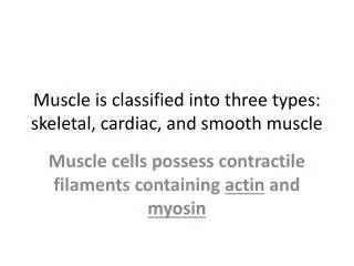
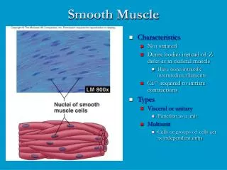
SMOOTH MUSCLE. Dr. Ayisha Qureshi MBBS, MPhil Assistant Professor. Structure of smooth muscle. STRUCTURE OF SMOOTH MUSCLE. Shape of muscle fiber: - spindle shaped - 1-5 µm in diameter - 20-500 µm in length

E N D
SMOOTH MUSCLE Dr. AyishaQureshi MBBS, MPhil Assistant Professor
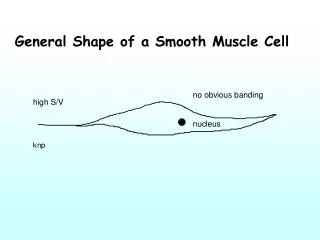
STRUCTURE OF SMOOTH MUSCLE • Shape of muscle fiber: - spindle shaped - 1-5 µm in diameter - 20-500 µm in length • A single nucleus present in the central thick portion. • Sarcolemma (cell membrane). • Cytoplasm appears homogenous without striations. • Fewer mitochondria as compared to the skeletal muscle. • Metabolism mostly glycolytic. • Actin, Myosin & Tropomyosin but NO Troponin
STRUCTURE OF SMOOTH MUSCLE • Dense bodies present attached to the cell membranes OR dispersed throughout the cell Dense bodies serve the same purpose as the Z-discs • Attached to the dense bodies are numerous numbers of Actin filaments • Interspersed between the actin filaments are Myosinfilaments ( their diameter twice as much as actin filaments) Usually, 5-10 times as many actin filaments as Myosin filaments
STRUCTURE OF SMOOTH MUSCLE • SIDEPOLAR CROSS-BRIDGES: Myosin filaments have sidepolar cross-bridges ↓ Bridges on one side hinge in one direction & on the other side in the opposite direction ↓ Allows myosin to pull an actin filament in one direction while simultaneously pulling it in the other direction on the other side ↓ Allow smooth muscle to contract 80% as compared to only 30 % in the skeletal muscle (force of contraction in skeletal muscle is limited because of the presence of the z-disc, against which the thick filament will abutt against and cannot move any further) • Calcium Pump: pumps Ca back into the SR if present for relaxation to take place. But it is very slow so that duration of cont. is longer.
STRUCTURE OF SMOOTH MUSCLE • Neuromuscular Junction: Does not occur in smooth m. Instead the autonomic nerves make diffuse junctions that secrete NT into the matrix coating of smooth m. a few micrometers away from the muscle fiber Also the axons supplying them do not have terminal buttons but varicosities on their terminal axons that contain the vesicles containing the NT • Neurotransmitter: Apart from Ach, norepinephrine can also be released Instead of synaptic clefts, smooth muscles have contact junctions
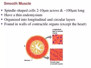
CLASSIFICATION OF SMOOTH MUSCLES UNITARY/ SINGLE UNIT/SYNCYTIAL/VISCERAL • Muscles of visceral organs .e.g. GIT, uterus, ureters & some of the smaller blood vessels. • Form a sheet or bundles of tissue. • Cell membranes show gap junctions that allows AP to pass rapidly from cell to cell. • AP spreads rapidly throughout the sheet of cells – cells contract as a single unit. MULTI-UNIT • Iris & Ciliary body of the eye, large arteries, Piloerector muscles • Showing discrete, individual smooth muscle fibers. • Smooth muscle cells not electrically linked. Each muscle fiber innervated by a single nerve ending. NT itself can spread and lead to an AP. • Selective activation of each muscle fiber that can then contract independently of each other.
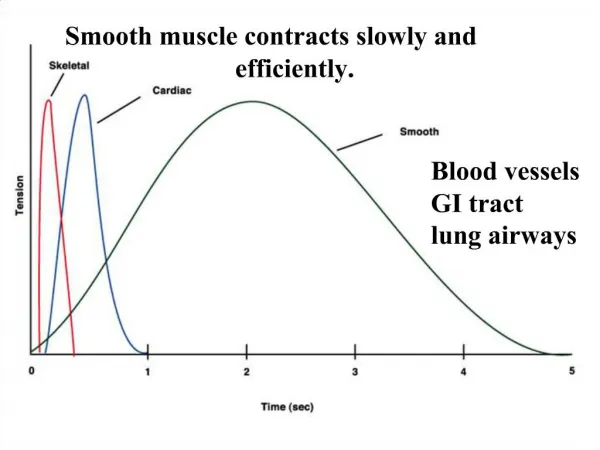
1. SINGLE MUSCLE TWITCH Single muscle contraction (muscle twitch) develops more slowly & relaxes even more slowly----Thus, longer sustained contraction without fatigue! Advantage:This ability allows the walls of the organs to maintain tension with a continued load .e.g. urinary bladder filled with urine A TYPICAL SMOOTH MUSCLE HAS A TOTAL CONTRACTION TIME OF 1-3 SECONDS (about 30 times as long as single skeletal muscle contraction)
2. ACTION POTENTIAL • In the normal resting state, the membrane potential is about -50 to -60 mv. • The AP of visceral smooth muscle is of 2 types: • Typical spike potentials: (similar to skeletal muscles) -mostly seen in the unitary smooth muscles • AP with Plateaus: Starts like a typical spike potential but repolarization delayed for several hundred to as many as 1000 msec ----accounts for the prolonged contraction that occurs in certain organs
2. ACTION POTENTIAL SLOW WAVE POTENTIALS: Without an external stimulus membrane potential is often associated with a basic slow wave rhythm. This is itself not an AP but a local property of the smooth muscle fibers. CAUSE: • Waxing & waning of the pumping of Na ions • Conductance of the ion channels increase & decrease rhythmically IMPORTANCE: When the peak of the slow wave reaches about -35 mv, threshold is reached and an AP develops & leads to a contraction. Thus, at peak of the slow waves an AP can occur. These slow waves are called as Pacemaker waves.
Action Potential Slow wave potentials Pacemaker potentials
3. ROLE OF CALCIUM • Poorly developed SR • Presence of caveolae- Small invaginations abut the SR which release Ca when AP reaches it. Thus, smooth muscle contraction is highly dependent on Extracellular Calcium conc. Point to Note: So the main source of Calcium ions in smooth muscle is to greater extent ECF and to a lesser extent SR as compared to the skeletal muscles where greatest source of Calcium is SR. Calcium plays the main role in the prolonged contraction process.
When unitary (visceral) smooth m. is stretched, spontaneous AP is usually generated, because: Normal slow potentials caused by stretch Overall ↓ in memb. Negativity caused by stretch
SMOOTH MUSCLE CONTRACTION SEQUENCE OF EVENTS: Binding of Ach to the receptors ↓ Increased Influx of Ca into the cell from the following sources: • ECF thru Ca channels • Ca released from SR • Stretch-activated Ca channels when memb. Deformed • Chemical-gated Ca channels by NT & hormones ↓ Ca binds to Calmodulin ↓ Ca-Calmodulin activates the enzyme: Myosin light chain kinase MLCK or simply Myosin kinase ↓ Phosphorylation of myosin, using energy & Pi from ATP ↓ Increased ATPase activity & binding of myosin to actin ↓ Contraction of smooth muscle
SMOOTH MUSCLE RELAXATIONSEQUENCE OF EVENTS: Dephosphorylation of Myosin by myosin phosphatase/ MLCP ↓ Decreases its ATP activity ↓ Ca removed from cytoplasm using Ca-Na antiport protein & Ca-ATPase ↓ Calmodulin releases Ca & uncomplexes from MK ↓ MK is phosphorylated by Protein kinase, inactivating it ↓ Relaxation OR sustained contraction
Latch system It is a state in which the dephosphrylated myosin remains attached to actin for prolonged period of time. This produces sustained contraction without consuming ATP & thus enables the smooth muscle to sustain long-term maintenance of tone without fatigue. E.g. urinary bladder full of urine.