Download
1 / 52
530 likes | 823 Views
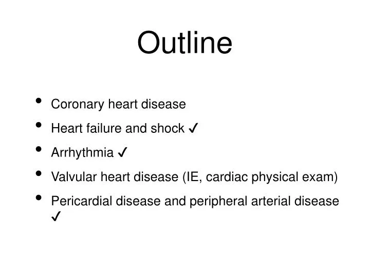
Outline. Coronary heart disease Heart failure and shock ✔ Arrhythmia ✔ Valvular heart disease (IE, cardiac physical exam) Pericardial disease and peripheral arterial disease ✔. Heart Failure. Heart failure, stage A, with diabetes Systolic heart failure, stage B, with old anteroseptal MI

E N D
Outline • Coronary heart disease • Heart failure and shock ✔ • Arrhythmia ✔ • Valvular heart disease (IE, cardiac physical exam) • Pericardial disease and peripheral arterial disease ✔
Heart Failure • Heart failure, stage A, with diabetes • Systolic heart failure, stage B, with old anteroseptal MI • Diastolic heart failure, NYHA Fc II, stage C, due to hypertensive heart disease • Systolic heart failure, NYHA Fc IV, stage D, due to dilated cardiomyopathy
Framingham Criteria for Heart Failure The diagnosis of heart failure in this study required that two major or one major and two minor criteria be present concurrently. Minor criteria were acceptable only if they could not be attributed to another medical condition. Minor Criteria Bilateral ankle edema Nocturnal cough Dyspnea on ordinary exertion Hepatomegaly Pleural effusion Tachycardia (rate ≥ 120 beats/min) Decrease in vital capacitiy by one third from maximal value recorded Major Criteria Paroxysmal nocturnal dyspnea Neck vein distention Rales Radiographic cardiomegaly Acute pulmonary edema S3 gallop Increased central venous pressure > 16 cm H2O Circulation time ≥ 25 sec Hepatojugular reflux Pulmonary edema, visceral congestion, or cardiomegaly at autopsy Weight loss ≥ 4.5Kg in 5 days in response to treatment of heart failure
Hemodynamic Model of HF BP = CO × SVR CO = SV × HR SV = ∫ (preload, contractility, afterload)
Causes of Heart Failure Impaired Cardiac Function Excess Work Demands Myocardial Disease Cardiomyopathies Myocarditis Coronary insufficiency Myocardial infarction Increased Pressure Work Systemic hypertension Pulmonary hypertension Coarctation of the aorta Valvular Heart Disease Stenotic valvular disease Regurgitant valvular disease Increased Volume Work Arteriovenous shunt Excessive administration of intravenous fluids Congenital Heart Disease Increased Perfusion Work Thyrotoxicosis Anemia Constrictive Pericarditis Carol Mattson Porth. Essentials of Pathophysiology. 2nd Ed
Management of Patients with HF • Acute decompensated HF • Chronic HF CO = SV × HR SV = ∫ (preload, contractility, afterload) * Removing precipitating causes* Control of congestive state Prevent cardiac remodeling Correction of underlying causes Prevent sudden cardiac death
Zipes: Braunwald's Heart Disease: A Textbook of Cardiovascular Medicine, 7th ed
Zipes: Braunwald's Heart Disease: A Textbook of Cardiovascular Medicine, 7th ed
Classification of Shock Circulatory shock Cardiogenic shock Hypovolemic Loss of whole blood Loss of plasma Loss of extracellular fluid Cardiogenic Myocardial failure Valvular failure Rhythm disorders Obstructive Inability of the heart to fill properly (cardiac tamponade) Obstruction to outflow from the heart (pulmonary embolism, cardiac myxoma, pneumothorax, or dissecting aneurysm) Distributive Loss of sympathetic vasomotor tone Presence of vasodilating substances in the blood (anaphylactic shock) Presence of inflammatory mediators (septic shock) in the blood Carol Mattson Porth. Essentials of Pathophysiology. 2nd Ed
Oxygen carrying capacity of Hgb: 1.39 SaO2 + 0.0031 PaO2 (mL/dL)
Atrial Fibrillation • Diagnosis • Etiology • Rate vs rhythm control? • Anticoagulation?
Causes of Atrial Fibrillaton Cardiovascular causes Non-cardiovascular causes Ischemic heart disease* Hypertension* Mitral valve disease Mitral stenosis* Mitral regurgitation Mitral valve prolapse Rheumatic heart disease Heart failure Cardiomyopathy Pericarditis Endocarditis Myocarditis Congenital heart disease Sinus node dysfunction Cardiac tumors Post-cardiac surgery Supraventricular arrhtyhmia Wolf-Parkinson-White syndrome Atrioventricular nodal re-entrant tarchycardia Metabolic Thyrotoxicosis* Low potassium, magnesium, or calcium; acidosis Pheochromocytoma Drugs (sympathomimitics) Alcohol (“holiday heart syndrome”) Postoperative (non-cardiac surgery) Hypothermia Respiratory causes Pneumonia Carcinoma of the lung Trauma Thoracic surgery Other causes Vagal atrial fibrillation Adrenergic atrial fibrillation Intracranial hemorrhage “Lone” atrial fibrillation *Most common causes
David H. Spodick: Pathophysiology of Cardiac Tamponade. CHEST 1998;113:1372-78
心包膜填充的臨床表現 • Beck’s triad 動脈血壓降低, 心音變弱, 頸靜脈壓升高 • Pulsus paradoxus 吸氣時收縮壓下降 >10mmHg
A 64M is brought into the ED with a chief complaint of substernal chest pressure for the past 30 minutes. This is associated with nausea and diaphoresis. He has a Hx of angina, which was previously relieved with NTG. He is a 30 pack year smoker. He has had three tablets of SL NTG but the pain persists. Physical exam reveals a heart rate of 80 bpm, resp of 30 bpm, and BP of 140/60 mmHg. The ECG demonstrates ST segment elevation in leads II, III, and aVF.1. Based on these symptoms and findings, which wall of the heart do suspect is affected?2. He is transferred to the CCU where thrombolytic therapy is initiated. His chest pain is relieved. An additional sign of successful reperfusion would be...
A 88M is admitted from the nursing home in acute CHF. Nursing home staff reports that his normal weight is 71Kg. Upon admission his vital signs include a heart rate of 104 bpm, resp of 28 bpm, and BP of 120/60 mmHg. His weight is 73.5Kg. Cardiovascular exam reveals regular heart rate and rhythm. There are crackles in both lung bases. after several hours in the ICU he becomes increasingly short of breath and you hear an S3 at the apex. He BP had decreased to 80/60 mmHg and his heart rate had risen to 130 bpm. He is breathing 40 times per minute. 1. Your immediate treatment goals are to...2. Which pharmacotherapeutic agents would you select to achieve these goals in this patient?3. Which of the following categories of medications are used in CHF patients to block a maladaptive compensatory mechanism?
When performing hypertension screening on a new patient you determine that the right arm BP is 150/98 mmHg. No other abnormalities are found. It is important to remember that:a. Hypertension may be diagnosed with as single elevated reading if it is in excess of 150/95 mmHgb. Hypertension is diagnosed after three consecutive readings >140/90 mmHgc. Hypertension of this magnitude requires institution of pharmacologic therapyd. Hypertension of this magnitude is of no concern and the patient can be assured
Which of the following classes of antihypertensive agents would be the best choice for a diabetic patient with hypertension?a. Calcium channel blockersb. Diureticsc. Beta-blockersd. ACE inhibitors
Risk factors for coronary heart disease in women include all of the following EXCEPT:a. A FHx of coronary heart diseaseb. Diabetesc. Obesityd. Hypothyroidism
A 55M with angina is currently taking beta-blockers and has moderate left ventricular dysfunction with an EF of 30%. You plan to supplement his medication regimen. Which drugs should be used cautiously in this patient?a. Transdermal nitrateb. Calcium channel blockersc. ACE-inhibitorsd. Angiotensin receptor blockers
Causes of secondary hypertension include all of the following EXCEPT:a. Renal artery stenosisb. Aortic aneurysmc. Cushing’s syndromed. Pheochromocytoma