Download
1 / 23
360 likes | 705 Views
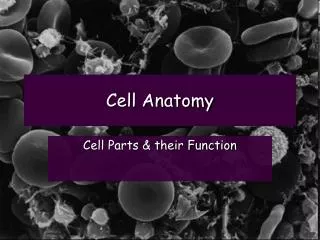
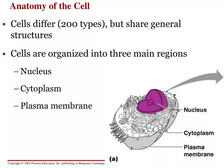
Anatomy of the Cell. Cells differ (200 types), but share general structures Cells are organized into three main regions Nucleus Cytoplasm Plasma membrane. Figure 3.1a. The Nucleus - Control center. Contains genetic material (DNA) Nuclear envelope w/ pores Nucleolus Chromatin.

E N D
Anatomy of the Cell • Cells differ (200 types), but share general structures • Cells are organized into three main regions • Nucleus • Cytoplasm • Plasma membrane Figure 3.1a
The Nucleus - Control center Contains genetic material (DNA) • Nuclear envelope w/ pores • Nucleolus • Chromatin Figure 3.1b
nucleus nuclear pores
Chromatin • Composed of DNA and protein • Scattered throughout the nucleus • Chromatin condenses to form chromosomes when the cell divides
Transport vesiclebuds off 4 Ribosome Secretory(glyco-) proteininside transportvesicle Sugarchain 3 Glycoprotein 1 2 ROUGH ER Polypeptide Rough endoplasmic reticulum • Makes proteins, membranes Figure 4.8
The Golgi complex finishes, sorts, and ships cell products Golgi apparatus Golgiapparatus “Receiving” side ofGolgi apparatus Transportvesiclefrom ER Newvesicleforming “Shipping”side of Golgiapparatus Transport vesiclefrom the Golgi Figure 4.10
Lysosomes • sacs of digestive enzymes • digest food. bacteria • recycle damaged organelles • embryonic development • waste storage Pombe’s disease - glycogen Tay-Sachs disease - lipids LYSOSOME Nucleus Figure 4.11A
Rough ER Transport vesicle(containing inactivehydrolytic enzymes) Plasmamembrane Golgiapparatus Engulfmentof particle Lysosomeengulfingdamagedorganelle “Food” LYSOSOMES Digestion Foodvacuole Figure 4.11B
Smooth endoplasmic reticulum • synthesizes lipids • regulates carbohydrate metabolism (liver) • breaks down toxins and drugs (liver) • Stores Ca++ in muscle cells
cellular respiration provides energy Mitochondrion Outermembrane Intermembranespace Innermembrane Cristae Matrix Figure 4.16
Cytoplasmic Organelles Figure 3.4
The CYTOSKELETON helps organize a cell’s structure and activities • network of protein fibers - microfilaments, microtubules Figure 4.17A
Cilia and flagella • appendages that protrude from certain cells • Function: movement • Made of microtubules wrapped in the plasma membrane • Centrioles - movement of chromosomes in cell division
Animal cells - surrounded by an extracellular matrix • sticky layer of glycoproteins • binds cells together in tissues • protects and support cells
Plasma Membrane Specializations • Microvilli • increase surface area for absorption • Membrane junctions • Tight junctions • Desmosomes • Gap junctions Figure 3.3
Cells and Tissues • Cells = building blocks of all living things • Carry out chemical activities needed for life • Tissues are groups of cells that are similar in structure and function