Download
1 / 49
631 likes | 3.86k Views
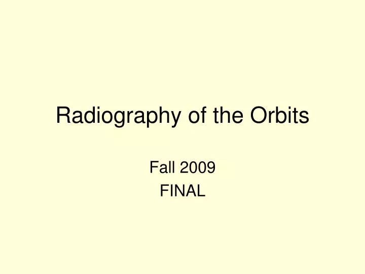
Radiography of the Orbits. Fall 2009 FINAL. Function of Orbits. Serve as bony sockets for the eyeballs Openings for nerves and blood vessels. Bones of the Orbits - 7. A. B. C. D. E. I. F. G. H. ________________ Primarily composed of orbital plate of frontal bone

E N D
Radiography of the Orbits Fall 2009 FINAL
Function of Orbits • Serve as bony sockets for the eyeballs • Openings for nerves and blood vessels
Bones of the Orbits - 7 A B C D E I F G H
________________ Primarily composed of orbital plate of frontal bone ________________ Zygoma (small amount) Maxilla Palatine 3. _______________ Medial Lacrimal Lateral Zygoma (large amount) Division of the Orbits
Base of the Orbit The circumference is made of 3 bones: • _________________ • _________________ • _________________
Openings in Posterior Orbit • ______________ • Optic canal • Sphenoid strut • ______________ • ______________
ANGLE OF ORBITS • Each orbit projects • 30 degrees superiorly • 37 degrees toward MSP
Indications for Orbit and Eye Radiography • Possible Fractures • Blowout • Tripod • Lefort • Foreign body of the eye
Mechanisms producing Orbital Fractures • Auto accidents • Assault • Falls, sports, and industrial accidents
Blowout Fracture • Blow to the eye • Orbital floor is fractured • Soft tissue herniates into maxillary sinuses • Often have ocular injury Ponsell, 2003
Tripod Fracture • Direct blow to zygoma • Visual concave abnormalities • Usually orbits are involved 4. Free floating zygoma Ponsell, 2003
LeFort Fractures LeFort types II & III involve the orbits Richardson, 2000
Type II Separation through: frontal processes lacrimal bones orbit floors, zygomaticomaxillary suture line lateral wall of maxillary sinuses pterygoid Complications ____________ system obstruction Infraorbital nerve anesthesia ____________ Malocclusion LeFort Fractures
Type III Separation of mid third of face at: zygomaticotemporal naso-frontal sutures and across orbital floors Complications _________________ Mal-union _________________ Lenthening of mid face _________________ system obstruction LeFort Fractures
Orbits Basic _________________ _________________ Special _________________ Eyes Basic _______________ _______________ _______________ Basic and Special Projections
Parietoacanthial ProjectionWaters Method • Positioning • ____________ • ____________ • Lines and planes: • ____________ • ____________ • CR: • ___________ Ballinger & Frank, 1999, pg 317
Parietoacanthial ProjectionWaters Method Radiograph • Distance from lateral border of skull and orbit equal on each side • Petrous ridges projected immediately below maxillary sinuses
Parietoacanthial Projection Modified Waters • Positioning: • _____________ • Lines and planes • _____________ • _____________ • CR • ____________
Modified Waters Radiographs • Petrous ridges projected immediately below the inferior border of the orbits • Equal distance from lateral orbit to lateral skull on both sides
Acanthioparietal ProjectionReverse Waters Method • Positioning • _______________ • Lines and planes • _______________ • _______________ • CR • _______________ Ballinger & Frank, 1999, pg 320
Reverse Waters Radiograph • Distance from lateral border of skull and orbit equal on each side • Petrous ridges projected immediately below maxillary sinuses
Parietoorbital Projection (Rhese Method)(PA) Optic Canal and Foramen • Positioning • ______________ • ______________ • Lines and planes: • ______________ • ______________ • PA- CR: • enters 1”superior and posterior to TEA • PA- CR: • exits through the affected orbit Ballinger & Frank, 1999, pg 290
Parietoorbital Projection (Rhese Method)(PA) Optic Canal and Foramen Radiograph • Optic canal & foramen visible at end of sphenoid ridge in inferior & lateral quadrant of orbit • Entire orbital rim • Supraorbital margins lying in same horizontal plane • Close beam restriction to the orbital region
Rhese Method(AP) Optic Canal and Foramen • Positioning: • _______________ • _______________ • Lines and planes: • _______________ • _______________ • AP- CR: • _______________ Ballinger & Frank, 1999, pg 292
Rhese Method(AP) Optic Canal and Foramen Radiograph • Optic canal & foramen visible at end of sphenoid ridge in inferior & lateral quadrant of orbit • Entire orbital rim • Supraorbital margins lying in same horizontal plane • Close beam restriction top the orbital region
Lateral Projection (EYE) • Positioning: • Semiprone or seated upright • Affected eye closest to cassette • ___________________ • Lines and planes: • MSP parallel • IPL perpendicular • CR: • ___________________
Lateral Eye for Foreign Body • Density & contrast permitting optimal visibility of orbit and foreign bodies • SI orbital roofs • Close beam restriction
PA Axial (EYE) • Positioning • Forehead & nose on IR. • Center IR ¾ “ distal to nasion • ________________ • Lines and planes: • ________________ • CR: • Through center of orbits, 30 degrees caudal
PA Axial Eye Radiograph • Petrous pyramids lying below orbital shadows • No rotation of cranium • Close beam restriction
Modified Waters (EYE) • Positioning: • IR at level of orbits • Rest pt’s chin on IR • Instruct pt to close eyes and hold eyes still • Lines and planes: • MSP perp • OML 50 degrees • CR: • Perp through mid-orbits
Modified Waters Radiograph • Petrous Pyramids lying well below orbital shadows • Symmetric visualization of orbits • Close beam restriction
References Ballinger, P.W. & Frank, E.D. (1999). Merrill’s atlas of radiographic positions and radiologic procedures. V2. New York: Mosby Ponsell, M.R. (2003). Assessing facial fractures in the emergency room. New Jersey Richardson, M.L. (2000). Facial and mandibular fractures. Retrieved May 5, 2007 from: http//www.rad.washington.edu/mskbook/facialfx.html