Download
1 / 24
260 likes | 776 Views
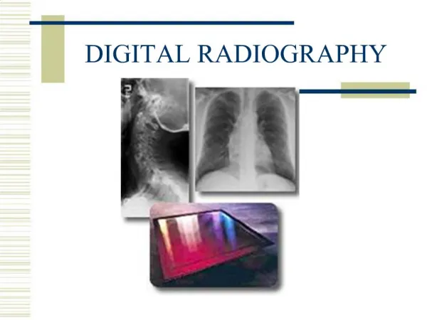
The Field of Digital Radiography. Loren Sachs Instructor. What is Digital Radiography?. Historically, digital radiography referred to specialized modalities that produced digital images. Examples would include: CT MRI Nuclear Medicine Ultrasound. Digital Radiography Today.

E N D
The Field of Digital Radiography Loren Sachs Instructor
What is Digital Radiography? • Historically, digital radiography referred to specialized modalities that produced digital images. • Examples would include: • CT • MRI • Nuclear Medicine • Ultrasound
Digital Radiography Today • Since the early 1990s, Digital Radiography has grown to include Computed Radiography(CR) and ‘true’ Digital Radiography(DR) or Direct Radiography. • Our lecture today will focus on CR and DR.
Digital Terms • Pixel • Picture element. This is the basic component of the digital image, it is what we see. 2 dimensional • Voxel • Volume element. This is a 3 dimensional element that includes depth. The pixel is essentially the end of the voxel.
Matrix • The actual image we see is made up of a series of pixels in rows and columns called a matrix. Generally, the larger the matrix the better the spatial resolution of the image. • Bit • The amount of gray scale in the image. The bit is the exponent to two, ie, a 2 bit image is two to the second or four. So the image would have 4 shades of gray possible. Today, most images are 10 or 12 bit.
FoV • Field of view. This is how much anatomy is displayed. A 12 cm FoV will display 12 cm of anatomy on the screen. The smaller the FoV the more magnified the anatomy is.
Window width • The gray scale of the digital image. The larger the width the more grays demonstrated the lower the contrast of the image. • Window level • The density or brightness of the image. The higher the number the brighter the image.
Computed Radiography • The technical aspects of CR are similar to what you see in the traditional analog radiology department, i.e., the technologist exposes a cassette that is then processed. • CR differs from analog in that the CR cassette contains a phosphor plate instead of a sheet of film.
The Technology of CR • After the cassette is exposed by the x-ray beam, the cassette is loaded into a reader. • The reader removes the phosphor plate and exposes it to a laser, stimulating the phosphors. • The light emitted from the plate is collected, quantified, and digitized.
Advantages of CR • The major advantage of CR is that existing radiology rooms can use the technology. • Consequently, the Radiology department can be digital at a relatively low cost, between $100,000-150,000 per reader. • Also, due to the similarities with traditional radiography the learning curve is much shorter with CR.
Digital Radiography • DR uses no cassette. The image capture device is embedded within the table-top. • The advantage here is that the steps involved in processing a ‘cassette’ are eliminated resulting in a huge increase in productivity. • Studies have estimated a 100% increase in room throughput.
Technology of Digital Radiography • There are two types currently being used • Direct • Indirect
Direct Digital Radiography The photoconductor is made up of amorphous selenium.
Indirect Digital Radiography The intensifying screen is made up of cesium-iodide crystals and the photodetector is made up of amorphous silicon.
Which DR Methodology is Best? • It depends entirely on who you listen to. • Basically, different manufacturers are using proprietary technology and claiming theirs is the best. • Ultimately, it will be decided by end-users.
DR Issues • The significant disadvantage for DR is cost. • Normally, rooms with DR technology have to be constructed from the ground up. Today, the average DR room cost is $400,000-600,000 plus construction costs. • Construction costs can be overwhelming, particularly in older buildings that need to be brought up to code.
How does CR and DR Affect You the Technologist? • RSV is less for both CR and DR when compared to film/screen combinations. Consequently, techniques are going to be higher. • Centering and collimation are REQUIRED. • You gain a tremendous amount of exposure latitude. • Post-processing manipulation allows you to adjust film quality.
How does CR and DR Affect You the Technologist? 2 • By selecting processing algorithms you can obtain different anatomical information with one exposure. • A PA chest film and be reprocessed as a PA rib film. • These methodologies are not a panacea for being a ‘bad’ technologist.
How does CR and DR Affect the Patient and Medical Care? • Patient dose goes up with these systems for an individual exam; however, there are fewer repeats. • Films are available hospital-wide almost immediately and can be view in multiple locations simultaneously. • No films get lost. • Everyone associated with a patient’s films becomes more efficient.
Other Considerations • In order to gain maximum efficiency with CR and DR a RIS/PACs systems needs to be deployed. • RIS, Radiology Information Systems, are used to manage non-image patient data, i.e. exam number, dictation status, and billing. • PACs, Picture Archiving Computer, allows for the distribution of images over a pre-described area.
Conclusion • Digital radiography will change the way technologists practice radiography. • However, it will not eliminate the need for a quality education and an understanding of radiology principles. In fact, digital radiography will require additional learning in order to maximize its usefulness.