Download

1 / 33
400 likes | 1.19k Views
RT 233. ORBITS. Parieto-acanthial (Waters) projection Modified Parieto-acanthial (shallow Waters) projection PA Axial (Caldwell) projection Lateral projection Acanthio -parietal (reverse Waters) projection Modified Acanthio -parietal(reverse shallow Waters) projection

E N D
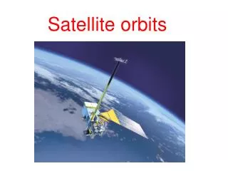
RT 233 ORBITS
Parieto-acanthial (Waters) projection • Modified Parieto-acanthial (shallow Waters) projection • PA Axial (Caldwell) projection • Lateral projection • Acanthio-parietal (reverse Waters) projection • Modified Acanthio-parietal(reverse shallow Waters) projection • Submentovertex for Zygomatic Arches • Submentovertex Tangential for Zygomatic Arches • Modified AP Axial (Townes) for Zygomatic Arches Facial bones projection review
Bones that make up the orbit • Frontal • Sphenoid • Ethmoid • Maxillary • Lacrimal • Palatine • Zygoma orbits
orbits • Circumference of orbits • 3 bones • Frontal bone • Maxiilla • Zygoma • Interior of orbits • 4 bones • Ethmoid • Sphenoid • Palatine • Lacrimal
Roof Primarily composed of orbital plate of frontal bone Floor Zygoma (small amount) Maxilla Palatine 3. Two Walls Medial Lacrimal Very thin and delicate Lateral Zygoma (large amount) Thickest wall Division of the Orbits
Openings in Posterior Orbit • Optic Foramen • Optic canal • Sphenoid strut • Superior Orbital Fissure • Inferior Orbital Fissure
Openings in the orbit Lateral view of the lateral orbital wall and its relationships. Key: FZS, frontozygomatic suture; FPZ, frontal process of the zygoma; ZB, zygomatic bone; ZA, zygomatic arch; ZFF, zygomaticofacial foramen; ZTF, zygomaticotemporal foramen; IOF, inferior orbital fissure; MS, maxillary sinus; TB, temporal bone; FB, frontal bone; GWS, greater wing of sphenoid.
ANGLE OF ORBITS • Each orbit projects • 30 degrees superiorly • 37 degrees toward MSP
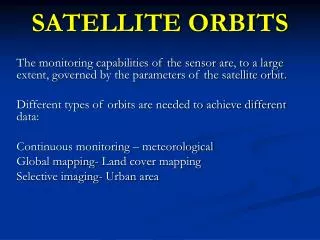
Orbital imaging • CT • MRI • Ultrasound • Plain film • Angiography Methods of imaging: • Possible Fractures • Blowout • Tripod • Lefort • Foreign body of the eye Indications for imaging:
Orbits Basic Parietoorbital (Rhese Method) Parietoacanthial (Waters method) Special Modified Parietoacanthial (Modified Waters method) PA Caldwell, (exag) Eyes Basic Lateral PA Axial Modified Parietoacanthial (Modified Waters method) Basic and Special Projections
Rhese Projection Rhese positioning
Orbits Series Waters orbital rims, zygoma, inferior orbital foramen Modified Waters (Shallow) inferior orbital rim in profile, Acanthioparietal same as Waters but magnified Rhese optic foramen PA Axial Caldwell superior sphenoid fissure Lateral Eye foreign body, soft tissue Waters foreign body, soft tissue Modified Waters foreign body, soft tissue PA Axial foreign body, soft tissue