Download
1 / 19
190 likes | 254 Views
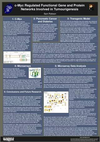
A Comparison of c-Myc Regulated Gene Networks Involved in Tumourigenesis of Two Distinct Tissues. Sam Robson MOAC DTC, Senate House, University of Warwick, Gibbet Hill Road, Coventry CV4 7AL. Cancer. Normal Mitosis. Abnormal Mitosis. = Healthy Cell. = Healthy Cell. = Cancer Cell.

E N D
A Comparison of c-MycRegulated Gene Networks Involved in Tumourigenesis of Two Distinct Tissues Sam Robson MOAC DTC, Senate House, University of Warwick, Gibbet Hill Road, Coventry CV4 7AL
Cancer Normal Mitosis Abnormal Mitosis = Healthy Cell = Healthy Cell = Cancer Cell = Cancer Cell = Apoptosis = Apoptosis Cell duplicates to form two identical daughter cells. Errors in DNA replication result in cell suicide (apoptosis) to avoid passing aberrant DNA to progeny. Genetic defects prevent apoptotic pathways from activating, allowing abnormal cells to proliferate. With no proliferative control, tumours can form.
C-Myc Legend Myc Box I Helix-Loop-Helix Leucine Zipper Myc Box II Basic Amino terminal Carboxyl terminal 1 45 63 129 355 368 410 439 143 C-Myc Protein Max Protein Transactivation domain Image adapted from Pelengaris et al. (2002).
Transgenic Model Legend Myc Box I Helix-Loop-Helix Leucine Zipper Myc Box II Basic Estrogen Receptor Max CACGTG TRRAP Myc-Max complex binds E-box sequence of target gene Transformation-Transcription domain Associated Protein (TRRAP) binds to MBII with help from MBI Inactive MycERTAM Active MycERTAM TRRAP recruits a histone acetyltransferase (HAT). This acetylates nucleosomal histones resulting in chromatin remodelling, allowing access by RNA Polymerase for gene transcription 4-Hydroxytamoxifen Myc HAT Max binds Myc at leucine helix-loop-helix zipper region RNA Polymerase 4-OHT binds estrogen receptor opening up bHLHz domain. Bound Heat Shock Protein 90 HSP90 ERTAM
Skin • Cells proliferate from epidermal stem cells in basal layer. • Migration towards surface – cells become keratinized. • Stratum corneus layer made up of highly keratinized nuclei-free cells – squames. • Squames constantly shed from epidermal surface. • Homeostasis within the skin very important. Image taken from http://kidshealth.org/kid/body/skin_noSW.html
Pancreas • Homogenous groupings of cells within the exocrine – Islets of Langerhans. • Islets contain predominately β-cells – sole source of insulin • Insulin responsible for glucose metabolism. • Loss of insulin leads to Type I diabetes. • Pancreatic ducts transport pancreatic enzymes. CACGTG CACGTG
c-MycERTAMActivation in Pancreas – Apoptosis CACGTG CACGTG INACTIVE ACTIVE +4OHT Apoptosis outweighs proliferation Islet involution Pancreatic Islet β-Cells Proliferation and Apoptosis c-MycERTAM Activation in Skin – Proliferation INACTIVE ACTIVE Cell Migration +4OHT Proliferation Increase in basal cell proliferation. More cells migrate through to squamous layer Skin epidermisConstant renewal of cells from basal layer. Keratinised ‘squame’ cells lost from surface Unchecked proliferation leads to many hallmarks of carcinoma. Tumour is localised with no metastasis seen.
Microarray • Features measure one nucleotide sequence (25mers). • Hundreds of identical 25mers per feature. • 11-20 features per gene. • 25mer sequence specifically binds biotin labelled cDNA. • Fluorescence readings give relative RNA concentration – equivalent to gene expression. Images courtesy of Affymetrix - www.affymetrix.com
AAAA B AAAA B AAAA B AAAA Microarray Hybridization Biotin-labelled cRNA Total RNA cDNA Reverse Transcription In Vitro Transcription Fragmentation, biotin labelling and hybridization. Analysis in Genespring
Laser Capture Microscopy LCM Ependorf Tube Membrane Slide Tissue Glass Slide Support Laser • Tissue section bound to membrane of LCM slide. Glass slide used as support on LCM platform. • “Sticky” ependorf tube lid lowered onto membrane. Laser cuts designated area for dissection. Raising lid lifts cut material from LCM platform for RNA extraction.
Laser Capture Microscopy 1: 2: Sweat gland Path of laser 3: 4: Sweat gland removed Laser captured sweat gland
Problems with pancreatic RNA • RNA degraded naturally in cells by the enzyme RNase. • Pancreas rife with RNase activity. • Integrity of RNA gradually decreases throughout LCM procedure. • RNA fully degraded by the time of tissue collection.
Exocrine RNA A Fresh Sample B Islet RNA C 18S and 28S peaks different heights 18S and 28S peaks different heights Large unknown peak Problems with pancreatic RNA • RNA integrity in islets is good compared to RNA integrity in exocrine tissue. • Implies that islet RNA is not subject to same degradation as that in the exocrine. • Possible that structure of islets protects islet cell RNA from ductal RNases.
Network Analysis • Empirical Bayesian approach estimates gene network structure from microarray data. • Problems – Sample size small for number of nodes (genes). • Allows estimation of gene interactions in complex system. Image from Schäfer et al., 2005.
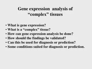
Generalised Linear Models • Unsupervised linear regression technique. • Models data as a linear combination of variables: • Gives the most statistically relevant variables. • Implementation in Genespring for public use. • Makes no assumptions of data and works with unbalanced experiments – Useful for clinical data.
Conclusion • c-Myc known to be very important in cancer formation. • c-Myc function in cancer onset still not fully understood. • In vivo analysis of early c-Myc activity will help to disentangle the web of c-Myc functionality. • Understanding of the route of tumourigenesis will hopefully aid in development of gene specific cancer therapies.
Acknowledgements Project Supervisors: Mike Khan David Epstein Stella Pelengaris Group members: Sylvie Abouna Linda Cheung Vicky Ifandi Göran Mattson Special thanks: Helen Bird, Sue Davis, Lesley Ward, David Pritlove, Sean James, Paul Anderson