Download
1 / 67
670 likes | 934 Views
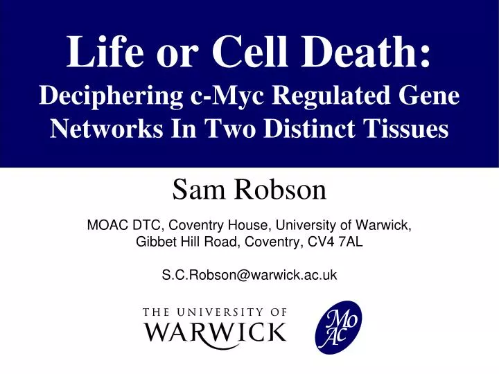
Life or Cell Death: Deciphering c- Myc Regulated Gene Networks In Two Distinct Tissues. Sam Robson MOAC DTC, Coventry House, University of Warwick, Gibbet Hill Road, Coventry, CV4 7AL S.C.Robson@warwick.ac.uk. Talk Outline. Introduction Skin vs. Pancreas Methods Quality Control Results

E N D
Life or Cell Death:Decipheringc-MycRegulated Gene Networks In Two Distinct Tissues Sam Robson MOAC DTC, Coventry House, University of Warwick, Gibbet Hill Road, Coventry, CV4 7AL S.C.Robson@warwick.ac.uk
Talk Outline • Introduction • Skin vs. Pancreas • Methods • Quality Control • Results • Linear Models • Conclusions
Project Aims • Analyse differences in gene-expression between two models of c-Myc activation with distinctly opposing phenotypes • Identify c-Myc targets that promote cell replication/survival and apoptotic cell death to help understand dual potential of c-Myc • To improve understanding of the complex activity of c-Myc in diseases such as cancer • To understand how c-Myc regulates vastly different paradoxical phenotypes in vivo
1: Introduction Transcription factor- wide range of cellular functions “Master regulator of genes” Deregulated in majority of human cancers Possible therapeutic target? We know WHAT c-Myc does, but we want to know WHY it does it
c-Myc Regulated Processes Angiogenesis Growth External Signals (eg. mitogens, survival factors) c-Myc Loss of Differentiation Proliferation Apoptosis
2: Skin vs. Pancreas Controlled activation of c-Myc in target cells of adult mice in vivo Targetted to pancreatic islet β-cells (insulin promoter) and skin supra-basal keratinocytes (involucrin promoter) Opposing phenotypic outcomes
c-MycERTAM Activation Inactive Active Suprabasal layer Skin Suprabasal layer Pancreas Pelengaris et al. (1999), Molecular Cell, Vol. 3(5), 565-577 Pelengaris et al. (2002), Cell, Vol. 109(3), 321-334
c-MycERTAM Activation Pelengaris et al. (1999), Molecular Cell, Vol. 3(5), 565-577 • SkinUnchecked proliferation, no apoptosis - Replication • PancreasSynchronous cell cycle entry and apoptosis – Death • Myc activation regulates two opposing phenotypes Pelengaris et al. (2002), Cell, Vol. 109(3), 321-334
3: Methods 2: Extraction of Tissue Excision of target tissue 3: Laser Capture Microdissection Isolation of homogenous tissue 1: Treatment of Transgenics Controlled activation of c-Myc in two diverse tissues QC QC 6: Microarray Hybridisation Hybridise fragmented, labelled cRNA to microarrays 5: 2-Cycle IVT Preparation of cRNA for microarray hybridisation 4: RNA Extraction Isolate total RNA from target cells QC 9: Functional Validation Linking results to the biology of the system 7: Microarray Data Analysis Analysis of microarray data 8: Validation Studies Validation studies to confirm results QC
Experimental Setup GeneExpression GeneExpression GeneExpression GeneExpression 4hr 4hr 4hr 4hr 8hr 8hr 8hr 8hr 16hr 16hr 16hr 16hr 32hr 32hr 32hr 32hr Myc OFF Myc ON n=3 n=3 Time course Time course Skin Tissue n=3 n=3 Time course Time course Pancreas Tissue
Laser Capture Microdissection β-cells make up only ~2% of pancreas Interesting changes masked by changes in non-Myc encoding cells LCM allows isolation of homogenous cell populations Nikon SL Microcut LCM system LCM Eppendorf Tube Laser Membrane Slide Tissue Glass Slide Support
Laser Capture Microdissection 1: Find Islet 2: Cut Islet 3: Lift Islet 4: Extracted Islet
Laser Capture Microdissection 1: Find Islet 2: Cut Islet 3: Lift Islet 4: Extracted Islet
Laser Capture Microdissection 1: Find Islet 2: Cut Islet 3: Lift Islet 4: Extracted Islet
18s and 28s peaks non-existent 18s and 28s peaks non-existent 18s and 28s peaks non-existent LCM Optimisation - Pancreas Control section • Pancreas has large number of RNases • RNA Integrity decreases rapidly using standard LCM protocols (Arcturus Pixcell, PALM MicroBeam, etc.) • RNA fully degraded before reaching laser capture platform
LCM Optimisation - Pancreas • All slides washed and baked before use • 100% ethanol used at all times • All surfaces cleaned thoroughly, gloves worn at all times, etc. • Don’t let tissue thaw (fix in ice-cold 100% ethanol) • Very quick staining (10 secs with 1% toluidine blue in 100% ethanol) • Air-dry sections in dessicator • Limit time on LCM platform (10 mins max)
LCM Optimisation - Skin • Difficult to cut – Strong cell-cell bonds • Slow laser burns and damages cells • Difficult to lift from surrounding tissue • Area of captured tissue much smaller than for islets • Difficult to obtain sufficient RNA yield • LCM not essential for skin compared to pancreas – decided to stick to whole sections
4: Quality Control • Quality control at every stage: • RNA extraction • IVT • Microarray hybridisation • Probe-level • Post-normalisation
Minimising Error • Technical Error • Same person performs all procedures • Meticulous planning • Standardise all protocols used • Randomisation of batches • Biological Error • Inbred transgenic mice used • All male • All same age • All culled at same time of day
Quality Control • Quality control at every stage: • RNA extraction • IVT • Microarray hybridisation • Probe-level • Post-normalisation
RNA Integrity RNA quality lower than threshold (RIN < 5)
Quality Control • Quality control at every stage: • RNA extraction • IVT • Microarray hybridisation • Probe-level • Post-normalisation
2nd Round cRNA Yield • Standard protocol results in low cRNA yield – Possible contaminant? • Using double volume vastly improves yield – Contaminant diluted?
2nd Round cRNA Yield Yield lower than recommended cutoff of 10 µg
Effect of RNA Quality on Yield General trend between RNA quality and 2nd round cRNA yield – Weakly correlated Low RIN does not necessarily mean poor yield High RIN samples can still give low yield RIN cannot accurately predict yield
Effect of RNA Quality on Yield Skin samples have higher RNA quality and yield than pancreas samples Many differences between skin and pancreas Greater ribonuclease activity in pancreas More intense processing for pancreas tissue RNA compared to skin
Quality Control • Quality control at every stage: • RNA extraction • IVT • Microarray hybridisation • Probe-level • Post-normalisation
Percent Present Low 2nd round cRNA yield (< 10 µg)
Scale Factor Low 2nd round cRNA yield (< 10 µg)
Quality Control • Quality control at every stage: • RNA extraction • IVT • Microarray hybridisation • Probe-level • Post-normalisation
Probe Level Models Example 1 Example 2 Example 3 • Pseudo-images of PLM summary - accounts for strong probe effects • Can see artefacts that may be otherwise hidden • Package affyPLM in Bioconductor in R .CEL File PLM ResidualsImage
Data Distribution – Pre-Normalised Pancreas Skin Myc OFF Myc ON
Quality Control • Quality control at every stage: • RNA extraction • IVT • Microarray hybridisation • Probe-level • Post-normalisation
Skin vs Pancreas Clustering – Group similar samples together Branching tree like structure – samples on the same branch most similar Data cluster nicely on tissue (some outliers) Given the protocol, the data looks great! Skin Pancreas Outliers
Data Distribution – Post-Normalised Pancreas Skin Myc OFF Myc ON
Removal of Outliers • Outliers tended to be poor across all QC tests • Good pancreas samples as outliers? • Remove or keep? Outliers to be removed
Effect of RNA Quality on Outliers • Outliers have wide range of RINs • Only one of four RIN < 5 samples classed as an outlier • Low RIN samples can produce good reproducible data
Effect of RNA Yield on Outliers • Outliers have wide range of 2nd round cRNA yields • Only 4 of the outliers had low cRNA yields • Good quality data with less than 4 µg cRNA!
5: Results • Early time point analysis – looking for direct effects of c-Myc activation • Untreated versus Treated • Combined 4 and 8 hour time points • Analysis in Genespring GX • Two-way ANOVA on tissue type and treatment, p = 0.05
6: Linear Models • Close collaboration with Agilent Technologies for linear modelling of microarray data • Part of multidisciplinary PhD - MOAC • Bioconductor package in R, GUI in Tcl/Tk • Implementation in GeneSpring GX (Agilent) • Simple to use for non-statisticians • Work in Progress - currently testing the program on a number of diverse data sets
Linear Models • Can be used in the following ways: • To ensure superfluous parameters have minimal effect on gene expression (eg batching effects) • To find interesting parameters • To find genes that change based on interesting parameters whilst taking other parameters and interactions into account (eg clinical data)
Linear Models vs ANOVA LM with 2 factors equivalent to 2-way ANOVA Comparable results to ANOVA Found a few genes (163) more than ANOVA for parameter 2 – borderline p-values Parameter 1 Interaction Parameter 2 LM ANOVA
7: Conclusions - What have I learnt? Talk to people (Affymetrix, UKAffy, GeneSpring Users Group, R/Bioconductor community, conferences, colleagues, etc.) Randomise everything Keep things really, really clean (be paranoid!) Plan everything with military precision Minimum of four replicates if possible You may still be able to get good results from poor quality RNA Using double volume of reagents in 1st cycle of IVT reaction can increase overall cRNA yield
Further Work • Analysis of microarray data • Use of LM tool and comparison of results with standard methods (ANOVA) • Validation of results – Immunohistochemistry, quantitative real time PCR, western blots, etc. • Functional validation – siRNA, ChIP-on-chip, etc. • Thesis…
Acknowledgements Project Supervisors:Michael KhanDavid EpsteinStella Pelengaris Collaborators:Helen BrownLesley WardSue DavisHeather Turner Ewan Hunter Special Thanks:Sheena LeePaul HeathGeoff ScopesGiorgia Riboldi-Tunnicliffe Sponsors: EPSRC, BBSRC, AICR, Eli Lilly and Amylin Pharmaceuticals Inc.