1 / 34

Figure 17.1 Apoptosis

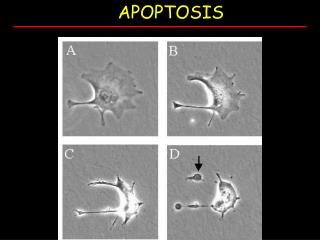
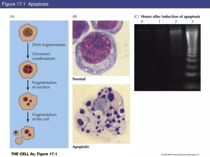
Figure 17.1 Apoptosis. Figure 17.2 Phagocytosis of apoptotic cells. Key Experiment 17.1: Photomicrographs of a normal worm (A) and a ced-3 mutant (B). Figure 17.3 Programmed cell death in C. elegans. Figure 17.4 Caspase targets. Figure 17.5 Caspase activation.

kearls
Download Presentation

Figure 17.1 Apoptosis

An Image/Link below is provided (as is) to download presentation Download Policy: Content on the Website is provided to you AS IS for your information and personal use and may not be sold / licensed / shared on other websites without getting consent from its author. Content is provided to you AS IS for your information and personal use only. Download presentation by click this link. While downloading, if for some reason you are not able to download a presentation, the publisher may have deleted the file from their server. During download, if you can't get a presentation, the file might be deleted by the publisher.

E N D

Presentation Transcript


  1. Figure 17.1 Apoptosis

  2. Figure 17.2 Phagocytosis of apoptotic cells

  3. Key Experiment 17.1: Photomicrographs of a normal worm (A) and a ced-3 mutant (B)

  4. Figure 17.3 Programmed cell death in C. elegans

  5. Figure 17.4 Caspase targets

  6. Figure 17.5 Caspase activation

  7. Figure 17.6 The Bcl-2 family

  8. Figure 17.7 Regulatory interactions between Bcl-2 family members

  9. Figure 17.8 The mitochondrial pathway of apoptosis

  10. Figure 17.9 Regulation of caspases by IAPs in Drosophila

  11. Figure 17.10 Role of p53 in DNA damage-induced apoptosis

  12. Figure 17.11 The PI 3-kinase pathway and cell survival

  13. Figure 17.12 Cell death receptors

  14. Figure 17.12 Cell death receptors (Part 1)

  15. Figure 17.12 Cell death receptors (Part 2)

  16. Figure 17.13 Skin fibroblasts

  17. Figure 17.14 Endothelial cells

  18. Figure 17.15 Proliferation of endothelial cells

  19. Figure 17.16 Liver regeneration

  20. Figure 17.17 Stem cell proliferation

  21. Figure 17.18 Formation of blood cells

  22. Figure 17.19 Renewal of the intestinal epithelium

  23. Figure 17.19 Renewal of the intestinal epithelium (Part 1)

  24. Figure 17.19 Renewal of the intestinal epithelium (Part 2)

  25. Figure 17.19 Renewal of the intestinal epithelium (Part 3)

  26. Figure 17.20 Stem cells of the skin

  27. Figure 17.21 Muscle satellite cells

  28. Figure 17.22 Hematopoietic stem cell transplantation

  29. Key Experiment 17.2: Embryonic stem cells differentiate in culture to a variety of cell types

  30. Figure 17.23 Culture of mammalian embryonic stem cells

  31. Figure 17.24 Differentiation of embryonic stem cells

  32. Figure 17.25 Cloning by somatic cell nuclear transfer

  33. Figure 17.26 Therapeutic cloning

  34. Figure 17.27 Induced pluripotent stem cells

More Related