Download
1 / 42
570 likes | 1.39k Views
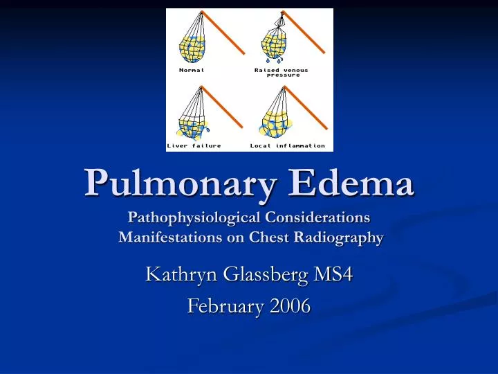
Pulmonary Edema Pathophysiological Considerations Manifestations on Chest Radiography. Kathryn Glassberg MS4 February 2006. Pulmonary Edema: Overview. Pathophysiology : Edema as an end result of a multitude of diverse insults (not just heart failure vs. ARDS!)

E N D
Pulmonary EdemaPathophysiological Considerations Manifestations on Chest Radiography Kathryn Glassberg MS4 February 2006
Pulmonary Edema: Overview • Pathophysiology : Edema as an end result of a multitude of diverse insults (not just heart failure vs. ARDS!) • Physiologic approach for radiologic evaluation of edema • Hydrostatic edema • Permeability edema +/- diffuse alveolar damage • Mixed permeability and hydrostatic edema
Pulmonary Edema • Edema occurs when physiologic resorption of fluid via lymphatics is overwhelmed • Causes usually divided into “hydrostatic” and “increased capillary permeability”, but both mechanisms can occur in the same patient! • Chest radiography, when combined with clinical data, helps distinguish pathologic cause in vast majority of cases
Pathophysiology overview2 Normally, excess hydrostatic transudate from pulmonary capillaries is filtered into peribronchovascular lymphatics and removed
Pathophysiology overview2 • In hydrostatic edema, transudate accumulates in the interstitum initially, only entering alveoli in severe cases • In permeability edema associated with diffuse alveolar damage (DAD), exudate fills the interstitum and the alveoli
Hydrostatic Edema3 • The lungs can accommodate increases in fluid: the lymphatic flow can increase 3-10x before edema develops • Higher hydrostatic pressures force fluid through endothelial cell pores, but the tighter junctions of epithelial cells prevent fluid from entering alveoli until pulmonary capillary pressures reach ~ 40 mm Hg, causing stress failure
Hydrostatic Edema: radiologic manifestations3 • Earliest sign: vascular indistinctness • Bronchial wall thickening/peribronchial cuffing • Septal lines: Kerley A, B, C • Thickened fissures • Severe edema: dependent ground glass opacities reflecting alveolar involvement • Often associated with bilateral transudative pleural effusions
Hydrostatic Edema: radiologic manifestations3 • “Cephalization” or “inversion” not specific for edema • Reflects chronic pulmonary venous changes in patients with left-sided heart failure • Vascular pedicle width • patients with volume overload often have widened vascular pedicles when compared to previous studies • However, patients can certainly have hydrostatic edema despite a narrow pedicle, thus this sign can be misleading
Vascular indistinctness Normal Edema Images courtesy of Dr. Marc Gosselin
Vascular Indistinctness Normal Edema Images courtesy of Dr. Marc Gosselin
Peribronchial cuffing • Images shown are pre- and post-treatment for hydrostatic edema • Arrowheads point to Kerley A lines
Septal Lines3 • The presence of septal lines reflects fluid accumulation between the lung lobules • Kerley lines • A: long, diagonal, central • B: short, horizontal, extend to lateral pleural surfaces • C: reticular pattern of ~ 1 cm polygons representing septal lines viewed on end (I’ve heard Dr. Kerley is the only one who has ever really seen these…)
Septal Lines Septal lines in a patient with cardiac failure
Septal Lines Lateral view of same patient– note fluid in both fissures
Septal Lines All three Kerleys claim to be present; can you find them?
Septal Lines Even in you can’t name the lines, you can see that this patient has severe hydrostatic edema in need of treatment! A B C?
Evolving hydrostatic edema4 33 year-old with AML admitted for renal failure and fluid overload
Arrows indicate peri-bronchial cuffing Note increasing size of azygous vein Evolving hydrostatic edema4
Evolving hydrostatic edema4 • Arrowheads indicate septal lines • Note ground-glass, indicating alveolar edema
Permeability Edema • multiple insults can cause increased pulmonary vessel permeability resulting in leakage of fluid AND protein • In its most severe form, the disease is a combination of vessel permeability and DAD, leading to the acute respiratory distress syndrome (ARDS)
ARDS pathology3 • Acutely, exudative edema in the alveoli causes hyaline membrane formation • Type II epithelial cells then proliferate and, usually, fibrosis occurs
ARDS: Radiologic manifestations3 • Patchy, diffuse ground glass opacities • Pattern of opacification does not change with position change, as the exudates are trapped in alveoli • Septal lines, peribronchial cuffing, and thick fissures are usually ABSENT • In severe cases, air bronchograms can be seen • Good rule of thumb: presence of ET tube!
ARDS: Radiologic manifestations3 Caution: While a normal sized heart and narrow vascular pedicle are helpful signs, neither is specific for injury edema
ARDS • Patchy diffuse ground glass • Air bronchograms • ET tube
Permeability Edema without DAD3 • Seen in IL-2 therapy for metastatic disease, hantavirus pulmonary syndrome • Severe capillary permeability without alveolar involvement • Radiographically, resembles hydrostatic edema (septal lines, peribronchial cuffing) because alveolar epithelium remains intact
Mixed hydrostatic and permeability edema • High-altitude pulmonary edema • Neurogenic edema • Reexpansion and post-obstructive
High-altitude pulmonary edema (HAPE)3 • Hypoxia causes non-uniform pulmonary vasoconstriction, leaving other lung units over-perfused and predisposed to edema • Higher pressures can result in some capillary damage and stress failure
High-altitude pulmonary edema3 • Radiographs show patchy ground glass with a central distribution favoring peribronchial cuffing and vascular indistinctness over septal lines
Neurogenic Edema3 • Pathophysiology similar to HAPE– neural mechanisms result in non-uniform vasoconstriction • High protein content of fluid indicates capillary leakage involved as well
Neurogenic Edema3 • Classically, neurogenic edema has an upper lobe predominance; however, it can present with any pattern • Often clears rapidly, arguing for intact alveoli
Neurogenic Edema4 • 54 year-old woman with intracranial hemorrhage • Note upper lobe predominance
Reexpansion and Postobstructive Edema3 • Both occur in setting of high negative pleural pressure • Reexpansion: usually seen as localized lung injury, with alveolar filling and exudative fluid, arguing for increased permeability as a cause • Postobstructive: pattern usually hydrostatic, secondary to increased central blood volume caused by the relief of obstruction
Reexpansion Edema4 Right pneumothorax One-hour post chest-tube placement
Postobstructive Edema4 Postextubation Laryngospasm: note central distribution and peribronchial cuffing.
Conclusions • Hydrostatic Edema is characterized by • Vascular indistinctness • Peribronchial cuffing • Septal lines/fissure thickening • Permeability Edema with DAD (ARDS) is characterized by • Diffuse, patchy ground glass opacities • Air bronchograms • Overlap is seen in pathophysiology, thus can be reflected in the radiograph
Hydrostatic and Permeability Edema Images courtesy of Dr. Marc Gosselin
“The condition of the capillary endothelium and that of the alveolar epithelium are the main determinants”3
References 1Milne ENC and Massimo P. Reading the Chest Radiograph: A Physiologic Approach. Mosby, 1993. 2Ware LB and Matthay MA. Acute pulmonary edema. The New England Journal of Medicine. 2005; 353: 2788-96. 3Ketai LH and Godwin JD. A new view of pulmonary edema and acute respiratory distress syndrome. Journal of Thoracic Imaging. 1998; 13: 147-171. 4Gluecker T. Capasso P. Schnyder P. Gudinchet F. Schaller MD. Revelly JP. Chiolero R. Vock P. Wicky S. Clinical and radiologic features of pulmonary edema.Radiographics. 19(6):1507-31; discussion 1532-3, 1999 Nov-Dec.
References • Images taken from: • myweb.lsbu.ac.uk/ ~dirt/museum/p6-71.html • www.bcm.edu/.../cases/ pediatric/text/7a-desc.htm • http://www.hcoa.org/hcoacme/chf-cme/chf00030.htm • http://www-medlib.med.utah.edu/WebPath/LUNGHTML/LUNG131.html • http://www-medlib.med.utah.edu/WebPath/LUNGHTML/LUNG133.html • http://www.lumen.luc.edu/lumen/MedEd/MEDICINE/PULMONAR/CXR/atlas/images/310a1.jpg • www.high-altitude-medicine.com/ AMS-medical.html • Sherman SC. Reexpansion pulmonary edema: a case report and review of the current literature. Journal of Emergency Medicine. Jan 2003; 24(1): 23-7. • Thanks to Dr. Marc Gosselin for images, insights