Download

1 / 27
280 likes | 510 Views
William 2001. Acute pulmonary edema. Causes: HF Permeability edema Both Most obstetric APE are due to noncardiogenic causes = 5% of ICU admissions = 0.5% of deliveries. Study: HF Preeclampsia Fluid overload Tocolytics Infection RF – HF

E N D
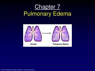
William 2001 Acute pulmonary edema
Causes: • HF • Permeability edema • Both Most obstetric APE are due to noncardiogenic causes = 5% of ICU admissions = 0.5% of deliveries
Study: • HF • Preeclampsia • Fluid overload • Tocolytics • Infection • RF – HF Tocolytics = 5% of obstetric causes
Commonly associated with: • Preeclampsia 28% • PTL 24% • Fetal surgery 17% • Infection 14% If iatrogenic causes are excluded, most cases are: • Old
Obese • Chronic HTN + superimposed preeclampsia Precipitation factors: • Operative delivery • acute blood loss • Anemia • Infection
Some causes of RF: - Pneumonia - Drug abuse - Sepsis - Arsenic poisoning - Hemorrhage - Pancreatitis - Preeclampsia - CT disease - Embolism - Pheochromocytoma - Irritant inhalation - Burns
= Worst form of RF Mortality: • Nonpregnant = 40 – 50% ( ↑ to 90% if + infection ) • Pregnant women = 25% Pathophysiological diagnosis include: • Alveolar epithelial injuries • Endothelial injuries Acute respiratory distress syndrome
Chemokines neutrophils recruitment ↑ cytokines tissue injury ↑ pulmonary capillary permeability ↓ lung volume ↑ arterial hypoxemia Criteria of diagnosis differ from: Mild pulmonary insufficiency to Total mechanical ventilation
Diagnosis: • X - ray diffuse infiltrates • PaO2 : FiO2 < 200 – 250 • No evidence of HF Most common cause: • Nonpregnant = sepsis • Pregnant = sepsis 40% = preeclampsia = hemorrhage
7% of cases are combination of: - Sepsis - Trauma - Shock - Fluid overload Clinical coarse depend on: • Magnitude of insult • Ability to compensate • Stage of disease
Very early: Hyperventilation Accentuation of pregnancy metabolic alkalosis + arterial O2 normal Later on: • X - ray and auscultatory evidence of lung disease • ↓ lung compliance
↑ Intrapulmonary blood shunting • Progressive alveolar and interstitial edema • Extravagation of WBCs and RBCs If not diagnosed RF: • Marked dyspnea • Marked tachypnea • Marked hypoxemia
With further ↓ of lung volume: • ↓↓ lung capacity • ↑↑ intrapulmonary blood shunting X–ray and chest auscultation Bilateral diffuse infiltrations Lethal if not treated with +ve airway pressure
Final phase: • ↑↑ intrapulmonary shunts ≥ 30% • Severe hypoxemia • ↑↑ dead space 60% of tidal volume • Hypercapnia ( = ↑ CO2 ) • Metabolic and respiratory acidosis • Myocardial irritability • HF
Histology of end stage: • Intra-alveolar fibrosis • Fibroblastic infiltration • Massive tissue plates Management: • O2 • Fluids/blood • Empirical antibiotics
Points: • O2α CO • Increasing PaO2 to 100 – 200 mmHg minimal ↑ of O2 delivery • Correction of anemia ↑↑ O2 delivery ( Each 1 gm of Hb carries 1.25 mL O2 when 90% saturated ) • Delivery does not improve hypoxia
Goals: • PaO2 = 60 mmHg • Or 90% oxyhemoglobin saturation • At an inspired O2 content of < 50% • With PEEP of < 15 mmHg Oxygen dissociation curve: Describes the propensity of Hb molecule to release O2
ODC is divided into: • Upper ODC represents alveolar - capillary environment Low O2 affinity high tissue capillary O2 exchange • Lower ODC represents tissue - capillary environment High O2 affinity PaO2 in maternal alveoli > tissue PaO2
With higher PaO2 in alveoli maternal Hb is maximally saturated Causes of right ODC shift: • Hypercapnia • Acidosis • ↓ temp • ↑ 2,3, diphosphglycerate level ( ↑ 30% during pregnancy ↑ O2 delivery to the mother and fetus )
Fetal Hb is characterized by: • ↑ O2 affinity • Left shift • Constantly in tissue portion of ODC - At any given PaO2 F Hb carries more O2 # M Hb - At high altitude maternal PaO2 = 60 mmHg while fetal PaO2 is at sea level
Mechanical ventilation: • In early stages O2 mask • In immanent RF intubation andartificial ventilation Adjustment of volume/cycle: • PaO2 ≥ 60 mmHg • PaCO2 35 – 45 mmHg • Hemoglobin saturation 90%
Positive-end-diastolic pressure: For severe pulmonary injury + highintrapulmonary shunting Filling of collapsed alveoli 5 – 15 mmHg no need for cardiovascular monitoring 15 mmHg ↓ VR ↓ CO ↓ uteroplacental circulation
Close PEEP during measuring PCWP higher results Other causes of high PCWP: • Overdistended alveoli • ↓ Compliance • Barotrauma Fluid therapy: Fluid overload worsen lung condition
Daily record: • Fluid intake/output • Body weight Mechanical ventilation add 1 L/day ↑ Permeability ↑ interstitial fluid Aim: Lowest PCWP possible + no ↓ CO Pregnancy changes: ↑ risk of lung injury from fluid therapy
Colloid oncotic pressure ( COP ): • Early during pregnancy = 28 mmHg • At term = 23 mmHg • During puerperium= 17 mmHg • Preeclampsia at term = 16 mmHg • During puerperium= 14 mmHg • COP/PCWP gradient = >8 mmHg • COP/PCWP gradient = ≤4 mmHg ↑ risk of pulmonary edema
Other therapy: • Surfactant • Nitric oxide • Corticosteroids • Immune therapy • Lipid mediator antagonists • Antioxidants