Download
1 / 35
430 likes | 1.25k Views
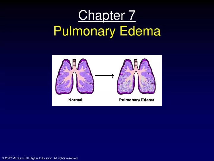
Chapter 7 Pulmonary Edema. Topics. Pulmonary edema Stages of Pulmonary edema Causes of Pulmonary edema Hypoxia caused by shunt Shunt measurement. Case Study #7: George. 55 yr old Stock Broker Well until 3 yrs ago Central chest discomfort during exertion

E N D
Topics • Pulmonary edema • Stages of Pulmonary edema • Causes of Pulmonary edema • Hypoxia caused by shunt • Shunt measurement
Case Study #7: George • 55 yr old Stock Broker • Well until 3 yrs ago • Central chest discomfort during exertion • Severe central chest pain on admission • Crushing pain which radiated to L shoulder • Very short of breath, coughed up frothy, clear fluid • 1 pack a day for 35 yrs • Father died at 60 of Heart attack
Physical exam #7: George • Anxious and SOB • Coughed up pink frothy fluid • BP 110/65, pulse 100 bpm • No neck vein engorgement • Rales upon ausculatation • No dependent edema • No edema
Investigations • ECG: recent L anterior wall infarct • Blotchy bilateral opacities in lungs • Po2= 59; Pco2= 35 mmHg; pH=7.35 • Lung scan (radioactive albumin): absent blood flow • Pulmonary edema and MI • Treatment: bed rest, morphine, diuretics and oxygen therapy • Pulm edema resolved quickly (2 days); sever obstruction of LAD CA; CABG performed
Pathophysiology • L.V. failure and pulmonary edema • Probably had CAD for several years • Pain on exertion was angina • The pain he felt on admission was from a myocardial infarction • When LV cannot function properly • Pressure builds up in the LA and also the Pulm. Veins • This increases Pulm capillary pressure, causes stress failure and alveolar flooding
Pathophysiology: Pulm Edema • Capillary endothelium is permeable to water and some solutes • Alveolar epithelium: • Much less permeable • Water actually travels in the other direction • Actively pumped from alveolus to interstitium (Na+-K+ ATP pump) • Hydrostatic forces: move fluid out of capillary • Osmotic forces: tend to oppose this
Physiology and Pathophysiology of the lung • Starling equation • Q=K[Pc-Pi)-σ(πc-πi)] • Q=net flow out of the capillary • K=filtration coefficient • Pc=cap hydrostatic pressure • Pi=interstitial pressure • πc=colloid osmotic press; cap • πi=colloid osmotic press; interstitium
Physiology and Pathophysiology of the lung • The values are not exactly know for most of the variables in the Starling eq • However, net flow out of cap at arterial end (higher Pcap) • Net inward flow and venular end; osmotic effect • Any excess outward fluid is collected in lymphatic vessels and returned to central circulation (empty into L subclavian v.)
Physiology and Pathophysiology of the lung • Note that in normal circumstances • Very little fluid build-up around either the vasculature or the bronchial tree • This increases in interstitial edema and reaches a critical stage in alveolar edema • Two factors limit the outward flow of fluid from caps • Colloid osmotic pressure is higher in the vasculature (and gets greater as mostly water is leaking out of caps) • Rise in hydrostatic pressure of interstitium as fluid passes out of caps
Interstitial and pulmonary edema • Interstitial edema • Engorgement of perivascular and peribronchial spaces (“cuffing”) • Pulm function minimally affected • Alveolar edema • Fluid in alveoli • Alveoli shrink (due to surface tension) • Ventilation is impaired • hypoxemia
Causes of pulmonary edema • Increased cap hydrostatic pressure • Recognized by measuring capillary “wedge” pressure (~pulm venous press.) • Increased cap permeability • Also inc. cap hydrostatic pressure • Reduced lymph drainage • Heart failure exacrebates this as central venous pressure rises • Decreased interstitial pressure • rare • Decreased colloid osmotic pressure • rare • Uncertain etiology • Heroin overdose
Features of pulm edema • Dyspnea • Rapid, shallow breathing • Stim of J receptors • Orthopnea • Paroxysmal nocturnal dyspnea • Periodic breathing • Cough • Pink, frothy discharge • Rales (crackles) • Rhonchi: musical sounds (severe edema) • Septal lines
Pulmonary function • Not normally done • Patients are pretty sick • Gas exchange • Impaired (particularly in alveolar edema) • Shunt • Blood that bypasses the gas exchange portion of lung; normally accounts for ~5mmHg A-a diff
Shunt • Shunt eq. • QT x CaO2 must equal • QS X CvO2 (shunt) and • (QT-QS) x CcapO2 • Rearranged as on Fig 7-6 • End result? • CaO2 is lower than optimal • Normally 1-2%
Shunt II • 100% does not correct hypoxemia due to shunt • Shunted blood never exposed to 100% O2 • This is actually HOW best to measure shunt • This is why O2concentration and Po2 are not raised very much by breathing 100% O2 • Actually rise in Cao2 is 0.003 ml/dl/mmHg Po2 • So, 600 x 0.003 = 1.8 so CaO2 rises only a small amount • Due to the flatness of the upper portion of the O2 dissociation curve
Hypoxemia • Pco2 does not rise with shunt; why? • Chemoreceptors • J receptors (George) • Hypoxemia (stim breathing) • Low VA/Q also contributes to hypoxemia • Obstructed airways are not ventilated • Low cardiac output also contributes to hypoxemia • In George; the CHF • Mixed venous Po2 falls due to increased O2 extraction by tissues
Pulmonary mechanics • Reduced distansibility of the lung • Reduces compliance • Airway resistance in increased • Some reflex VC • Some due to cuffing • Reduces the radial traction effect of increasing lung volume
Surface tension • Pressure is determined by the law of Laplace • P=4T/r • Thus, Pressure will fall as the radius of the sphere increases • Thus, a smaller sphere should empty into a larger sphere
Surface tension • Note that air inflation curve is right shifted • Reduced compliance • Due to surface tension • Surfactant • Reduces surface tension • Produced by type II alveolar cells • Contains a phospholipid; dipalmitoyl phosphatidylcholine (DPPC) • May be important contributor to respiratory distress syndrome in newborns
Surface tension • Main points • Water has very high surface tension • Placing detergent in water reduces surface tension • Lung extracts have variable surface tension • How/Why does surfactant work? • DPPC molecules are hydrophobic at one end and hydrophilic at the other; thus the individual molecules tend to repel each other, an effect that gets stronger as they get closer
Physiological advantages of surfactant • Low surface tension increases compliance • Stability of alveoli is improved (remember the tendency of small bubbles to empty into larger ones) • Surfactant reduces surface tension more in smaller bubbles • Keeps alveoli dry; elevated surface tension tends to suck fluids out of the low pressure caps
Other physiological changes with Pulmonary Embolism • Gas Exchange • Reduced Po2 • Atelectatic areas act as shunt • Pulmonary edema • Lung mechanics • Post-embolism areas receive no blood flow • Causes bronchoconstriction (reason why X-ray showed no ventilation in the region distal to emboli); short-lived usu.
Chapter 8: Pneumoconiosis • Or • Pneumonoultramicroscopicsilicovolcanoconiosis
Black lung disease: Harry • 60 yr old retired coal miner • SOB • Fatigue • Productive cough • Started working in mines at 17 • SOB started ~12 yrs ago • Smoked 1-2 packs a day since 15
Harry • Physical exam • Only positive findings • Chest slightly overinflated • Rhonchi heard on auscultation
Investigations • Blood work normal • X-ray showed fine particulate matter in lungs • Slightly elevated lung volumes • Slight obstruction and flow limitation • Slight hypoxemia
Pathogenesis • Types of pollutants • Carbon monoxide • Largest pollutant • Nitrogen oxides • Produced from burning of fossil fuels, like coal and oil (forms smog) • Sulfur oxides • Gases that come from burning sulfur containing fuels (power stations) • Hydrocarbons • Normally found in air; in combo with sunlight can cause Photochemical oxidants
Pathogenesis • Particulate matter • Wide variety of particles; soot • Power stations and industrial plants • Photochemical oxidants • Ozone, peroxy-nitrates • Formed from the action of sunlight on hydrocarbons and nitrogen oxides • Cigarette smoke • ~4% CO • Nicotine • Hydrocarbons (tar); causes bronchial carcinoma
Deposition of aerosols in the lung • Aerosol: particles that remain airborne for a substantial period of time • Impaction: • Largest particles fail to turn corners • Lodge in nasopharynx • Nose filters large particles well 5-20 μ are almost completely filtered
Deposition of aerosols in the lung • Sedimentation • Gradual settling of particles because of their weight • Medium sized particles • 1-5 μ; in small airways • Diffusion • Random movement of gases • Only in smallest particles; <0.1 μ • Many particles are exhaled; to small to sediment, to large to diffuse into blood
Clearance of deposited particles • Mucociliary clearance • Mucus: bronchial seromucus glands • Goblet cells • Film is 5-10 μ thick • Sol and gel layer • Gel: superficial, viscous; traps deposited particles • Sol: less viscous; allows beating of cilia
Clearance of deposited particles • Mucus Contains IgA • Important in defense against foreign particles, bacteria and viruses • Cilia: 5-7 μ long, beat 1000-1500 times/min • Propel gel layer forward • Moves at 1mm-2cm/min dependent upon the diameter of the airway • Eventually swallowed or “gobbed” up • Normal mucociliary clearance is impaired • Pollution • Toxic gases • Tobacco smoke
Alveolar macrophages • No mucociliary apparatus in alveoli • Macrophages • Engulf foreign particles via amoeboid motion • Phagocytose these particles (kill through lysozomal activity) • Can migrate to small airways and climb The mucociliary ladder • Leave blood in lymphatics (or blood) • Activity of macrophages is impaired by • Cigarette smoke, ozone, hypoxia, radiation, corticosteroids and alcohol
Other pneumoconioses • Coal worker’s lung • Massive fibrosis • Silicosis • Inhalation of silica • Quarrying, mining or snadblasting • These are toxic particles • Provoke severe fibrosis • Asbestos-related disease • Commonly used in insulation, brake linings, roofing materials (anything that must resist heat • Diffuse interstitial pulm fibrosis (Chpt 5) • Bronchial carcinoma; aggravated by smoking • Pleural disease; malignant mesothelioma (sometimes up to 40 yrs after exposure) • Byssinosis • Cotton dust • Histamine reaction • Obstructive disease pattern • Occupational asthma • Allergenic organic dusts • Flour; wheat weevil • Gum acacia • Polyurethane; Toluene diisocyanate