Download
1 / 16
160 likes | 276 Views
What if we want to know what allele(s ) of beta- globin an individual has?. Fig. 17-22. Wild-type hemoglobin DNA. Mutant hemoglobin DNA. C. T. T. C. 3 . 3 . A. T. 5 . 5 . T. 5 . A. G. G. A. A. 3 . 5 . 3 . mRNA. mRNA. G. A. A. 5 . G. U. A. 3 . 5 .

E N D
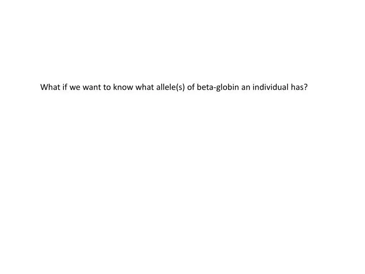
What if we want to know what allele(s) of beta-globin an individual has?
Fig. 17-22 Wild-type hemoglobin DNA Mutant hemoglobin DNA C T T C 3 3 A T 5 5 T 5 A G G A A 3 5 3 mRNA mRNA G A A 5 G U A 3 5 3 Normal hemoglobin Sickle-cell hemoglobin Val Glu
Fig. 17-22 Wild-type hemoglobin DNA Mutant hemoglobin DNA C T T C 3 3 A T 5 5 T 5 A G G A A 3 5 3 mRNA mRNA G A A 5 G U A 3 5 3 Normal hemoglobin Sickle-cell hemoglobin Val Glu DdeI cuts: CTNAG
Normal -globin allele Normalallele Sickle-cellallele Fig. 20-10 175 bp Large fragment 201 bp DdeI DdeI DdeI DdeI Largefragment Sickle-cell mutant -globin allele 376 bp 201 bp175 bp Large fragment 376 bp DdeI DdeI DdeI (a) DdeI restriction sites in normal and sickle-cell alleles of -globin gene (b) Electrophoresis of restriction fragments from normal and sickle-cell alleles
TECHNIQUE Heavyweight Restrictionfragments I II III Nitrocellulosemembrane (blot) DNA + restriction enzyme Gel Sponge I Normalinsulinallele II mutant insulinallele III Heterozygote Papertowels Fig. 20-11 Alkalinesolution 2 1 3 Preparation of restriction fragments DNA transfer (blotting) Gel electrophoresis Radioactively labeledprobe for insulin gene Probe base-pairswith fragments I II III I II III Fragment frommutant Insulin allele Film overblot Fragments fromnormal insulinallele Nitrocellulose blot 4 5 Probe detection Hybridization with radioactive probe
TECHNIQUE Heavyweight Restrictionfragments I II III Nitrocellulosemembrane (blot) DNA + restriction enzyme Gel Sponge I Normalinsulinallele II mutant insulinallele III Heterozygote Papertowels Fig. 20-11 Alkalinesolution 2 1 3 Preparation of restriction fragments DNA transfer (blotting) Gel electrophoresis Radioactively labeledprobe for insulin gene Probe base-pairswith fragments I II III I II III Fragment frommutant Insulin allele Film overblot Fragments fromnormal insulinallele Nitrocellulose blot 4 5 Probe detection Hybridization with radioactive probe
Another option: PCR of Beta-globin gene, followed by DdeI digest Normal -globin allele Normalallele Sickle-cellallele Fig. 20-10 175 bp Large fragment 201 bp DdeI DdeI DdeI DdeI Largefragment Sickle-cell mutant -globin allele 376 bp 201 bp175 bp Large fragment 376 bp DdeI DdeI DdeI (a) DdeI restriction sites in normal and sickle-cell alleles of -globin gene (b) Electrophoresis of restriction fragments from normal and sickle-cell alleles
How can we measure gene expression? wild type dif1 vs. Isolate RNA Compare gene expression
Reverse Transcriptase PCR (RT-PCR) TECHNIQUE 1 cDNA synthesis mRNAs cDNAs Fig. 20-13 Primers 2 PCR amplification -globingene 3 Gel electrophoresis Embryonic stages RESULTS 1 2 3 4 5 6
Where in the organism is my gene transcribed? Promoter : reporter fusions 50 µm
Where in the organism is my mRNA present? In situ hybridization Fig. 20-14 50 µm
TECHNIQUE Tissue sample 1 Isolate mRNA. 2 Make cDNA by reversetranscription, usingfluorescently labelednucleotides. mRNA molecules Fig. 20-15 Labeled cDNA molecules(single strands) DNA fragmentsrepresentingspecific genes 3 Apply the cDNA mixture to amicroarray, a different gene ineach spot. The cDNA hybridizeswith any complementary DNA onthe microarray. DNA microarray DNA microarraywith 2,400human genes 4 Rinse off excess cDNA; scanmicroarray for fluorescence.Each fluorescent spot represents agene expressed in the tissue sample.
Example of array data genes WT dif1 ∆ dif1 myb98 ∆ myb98
Large scale sequencing of cDNA fragments Reverse Transcriptase PCR (RT-PCR) TECHNIQUE 1 cDNA synthesis mRNAs cDNAs Primers 2 PCR amplification Sequence large numbers (millions) of cDNA fragments 3 Gel electrophoresis
Large scale sequencing of cDNA fragments No UV (3 samples) UV (3 samples) Fragments matching rad51