Download
1 / 37
390 likes | 822 Views
Applied Sciences Lecture Course. Ventilatory Failure & Hypoxia. Mahesh Nirmalan MD, FRCA, PhD Consultant, Critical Care Medicine Manchester Royal Infirmary. Objectives. Respiratory failure is one of the commonest manifestations of acute illness Hypoxia and CO 2 retention

E N D
Applied Sciences Lecture Course Ventilatory Failure & Hypoxia Mahesh Nirmalan MD, FRCA, PhD Consultant, Critical Care Medicine Manchester Royal Infirmary
Objectives • Respiratory failure is one of the commonest manifestations of acute illness • Hypoxia and CO2 retention • Failure of oxygen transfer • Failure of effective alveolar ventilation • Pathophysiology • Differences in management approach
Ventilation Moving an adequate volume of air Minute ventilation Alveolar ventilation Oxygenation Transfer of O2 across the alveoli Dusky colour Cyanosis Low SpO2 Low arterial PaO2 Respiratory rate Tidal volume or chest expansion Arterial PaCO2 Respiration or Breathing
Type 2 Type 1 Mixed Hypoxia & Hypercarbia Hypercarbia PaCO2>7kPa Hypoxia PaO2<8kPa Respiratory Failure
Treatment of Respiratory failure • Type 1 • Cause • O2 supplementation • PEEP • Type 2 • Cause • Ventilatory assistance • Pharmacological • Mechanical: IPPV • Mixed
Lung volumes FRC is a balance between two forces Reduced compliance Reduced FRC Increased compliance Increase in FRC
Inflammation and oedema within the lung parenchyma Low compliance and low FRC
Hepatisation Fibrinous exudate H’ge
Hyaline.membrane Normal lung Interstitial oedema Organising oedema Alveolar oedema Haemorrhage Neutrophil infiltartion Histological changes: reduced lung compliance
Reduced compliance • Pulmonary oedema • Pneumonia • ARDS and ALI • Fibrosis Tachypnoea Increased work of breathing Hypoxia
Decreased lung compliance • Tendency for the alveoli to collapse • May involve large parts of the lung • Reduction in FRC is an important factor • Increased work of breathing • Common cause for failure in oxygenation • Type 1 respiratory failure
Loss of elastic tissue within the lung parenchyma Increased lung compliance
Increased lung compliance: Increased FRC Hyper-inflation Low set diaphram Reduced lung markings
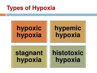
Hypoxia: failure of tissue oxygenation • Hypoxic hypoxia: Pulmonary oxygen transfer • Stagnant hypoxia: Poor blood flow • Anaemic hypoxia: poor oxygen carriage • Histotoxic hypoxia: Sepsis, Cyanide
Oxygen cascade in an ideal lung Diffusion, shunt, ventilation perfusion mismatch High Altitude Hypoventilation
CO2 retention: Ventilatory failure Treat the cause: Opiates, pain, airway obstruction Ventilatory support: Non-Invasive: BiPAP Invasive: Mechanical ventilation OXYGENATION: HYPOXIA Treat the cause: Infection, oedema ↑FiO2 PEEP Treatment of respiratory failure
Summary Failure of oxygenation • Hypoventilation • Diffusion • Shunt and V/Q mismatch • Treat the cause • Supplemental oxygenation & PEEP Failure of ventilation • Respiratory depression • Increase in physiological dead space • Treat the cause • Ventilatory assistance
Pathophysiology of hypoxia Venous blood Oxygenated blood
Pathophysiology of hypoxia Venous blood Venous blood
Ventilation/perfusion or V/Q mismatch Partially oxygenated blood Venous blood
Shunt and V/Q mismatch Alveolar oedema Shunt: blood that goes through unventilated lung units V/Q mismatch: Blood going through poorly ventilated units
Causes of Hypoxia Clinically how does one distinguish between shunt and V/Q mismatch? Effect of increasing FiO2 Hypoventilation Diffusion defects Ventilation-perfusion mismatch Shunts
45 years old male: Breathless, pyrexial, unwell, (breathing 50% O2)Pulse oximetry: 90% saturation • pH=:7.15 • PCO2: 3.3 kPa • PO2: 13.47kPa • HCO3-: 17 mmol.l-1 • Hb: 10.8 g.dl-1 • Glucose: 12.8mmol.l-1 • Lactate: 0.9mmol.l-1 Shunt and V/Q mismatch
Physiological dead space Wasted ventilation Extension of dead space Ventilated but not perfused alveolar units Physiological dead space Dead space ventilation does not clear CO2 Extension of dead space will lead to CO2 retention
Pulmonary embolism: Typically increase in Physiological dead space When large also causes significant V/Q mismatch Hypoxia and CO2 retention
Most organic parenchymal diseases:Increase in V/QSome shuntingIncrease in physiological dead space
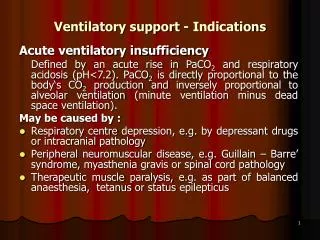
Ventilatory Failure • Hypoventilation • Depression of respiratory centre: opiates • Pain: upper abdominal surgery, Rib fractures • Prolonged increase in work of breathing • Tachypnoea • Reduced lung compliance • Severe asthma • Extension of physiological dead space • COPD
COPD: 25% O2 pH=:7.15 PCO2: 12.3 kPa PO2: 13.47 kPa HCO3-: 32mmol.l-1 Hb: 18.8 g.dl-1 Glucose: 9.8mmol.l-1 Lactate: 0.9mmol.l-1