Download
1 / 30
330 likes | 976 Views
HYPOXIA. Maroun Matta, M.D. Plan. I. Dyspnea v/s Hypoxia II. Mechanisms of hypoxia III. Evaluation of hypoxia A. Questions to ask over the phone B. Looking up the patient C. At bedside IV. ABG V. CXR and the differential VI. Treatment. I. Dyspnea v/s Hypoxia. I. Dyspnea v/s Hypoxia.

E N D
HYPOXIA Maroun Matta, M.D.
Plan • I. Dyspnea v/s Hypoxia • II. Mechanisms of hypoxia • III. Evaluation of hypoxia • A. Questions to ask over the phone • B. Looking up the patient • C. At bedside • IV. ABG • V. CXR and the differential • VI. Treatment
I. Dyspnea v/s Hypoxia • A. Dyspnea: “subjective” sensation, not always associated with hypoxia. • B. Hypoxia: low SaO2 and PaO2 (no data to support an exact number) by convention: • SaO2 less than 95% (doesn’t work for everyone!) • PaO2 less than 80 mmHg
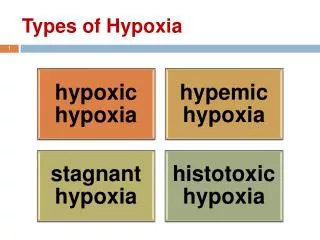
II. Mechanisms of hypoxia • A. Reduced inspired oxygen (Altitude) • B. Diffusion limitation (severe ILD, very rare) • C. Hypoventilation (Narcotics, obesity and hypoventilation sd, CNS injury, Neuro-muscular disease) • D. V/Q mismatch • E. R-L shunt (v=0)
III. Evaluation of hypoxia • Nurse Calls about Mr X, SaO2 91% What questions should ask over the phone? (7 questions!)
A. Questions over the phone • 1) on how much oxygen? • 2) what is his baseline oxygen requirement? • 3) Heart Rate (looking for arrythmia order ECG) • 4) BP (hypertension emergency and falsh pulmonary edema) NB: hypercapnia can cause increase HR and BP • 5) Fever (infection, clot) • 6) I/O for the last 24-48hr (weight?!) • 7) Is the patient complaining of any sob, cp, palpitations? Is his mental status altered?
What information to look for when looking up the patient or your way to his room?
B. Patient’s Background • 1) Age/Sex • 2) PMH (lung/Heart disease) • 3) Admission: date, service, reason for admission • 4) Recent procedures ( to suggest pneumoth.) • 5) Medications (inhalers, diuretics, narcotics and dvt prophylaxis) • 6) Labs (new leucocytosis, bnp) • 7) Imaging (last echo, cxr) • 8) CODE STATUS !!!
C. At Bedside • 1) Looking at the patient • 2) Pertinent hx • 3) key PE findings
1. Looking at the patient • A) work of breathing (labored v/s relaxed) • B) signs of cyanosis • C) signs of hypercapnia (flushed skin, diaphoresis)
2. Pertinent Hx • FIRST note patient’s speech (words v/s full sentences) • 1) Mentation • 2) sob, cough, sputum • 3) chest pain, palpitations, orthopnea • 4) For how long? Is it the first time?
3. Pertinent PE • 1) JVP • 2) Lung auscultation (stridor, wheezing, crackles, absent breath sounds uni/bila) • 3) Heart auscultation (tachy/regular, s3, s4, murmurs, distant?) • 4)LE (edema, dvt signs)
IV. Value of an ABG very limited in acute settings! • 1) SaO2 correlates very nicely w PaO2 (exceptions hypothermia, shock, CO poisoning, methemoglobinemia) • 2) PH and Bicarbonate/PCO2 correlates on VBG correlates very nicely with ABG • NB: chronic settings A-a gradiant.
V. CXR and differential • A) Normal (PE until proven otherwise!) • B) Abnormal • 1) Diffuse • A) interstitial ( fluids v/s blood) • B) alveolar (inflammation ARDS) • 2) Focal • A) infiltrate (pna) • B) effusion (pleural effusion) • C) pneumo/hemothorax
VI. Management • 1) Assure Oxygenation/Ventilation • 2) Inhalers • 3) Diuresis • 4) Antibiotics • 5) Heparin drip • 6) MICU transfer
1) Oxygenation / Ventilation A) Oxygenation NC, VN, NR, CPAP/BiPaP, Intubation B) Ventilation BiPap, Intubation
2) Inhalers • Asthma exacerbation (clear cxr, hx of asthma, wheezing on exam) • COPD exacerbation (variable cxr, hx of copd, w increased cough and sputum)
3) Diuresis • Volume Overload (increased intersitial markings, HF patient, w elevated jvp, may cause wheezing!, keep in mind that crackles and lee are very advanced findings) • Flash pulmonary edema (control bp)
4) Antibiotics • PNA (new infiltrate, w leucocytosis, fever, increased cough and sputum)
5) Heparin Drip • PE (wills score, cxr normal or UNCHANGED)
6) MICU transfer • Patient is going to require invasive ventilation