Download
1 / 27
270 likes | 274 Views
Determinants of Cardiac Output and Principles of Oxygen Delivery. Scott V Perryman, MD PGY-III. Principle of Continuity: Conservation of mass in a closed hydraulic system Blood is an incompressible fluid Vascular system is a closed hydraulic loop

E N D
Determinants of Cardiac Output and Principles of Oxygen Delivery Scott V Perryman, MD PGY-III
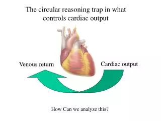
Principle of Continuity: • Conservation of mass in a closed hydraulic system • Blood is an incompressible fluid • Vascular system is a closed hydraulic loop • Vol ejected from left heart = vol received in R heart
Preload • Preload: load imposed on a muscle before the onset of contraction • Muscle stretches to new length • Stretch in cardiac muscle determined by end diastolic volume
Preload • At bedside, use EDP as surrogate for ventricular preload • i.e. assume EDV = EDP
Preload • How can we measure EDP? Pulmonary Capillary Wedge Pressure
PCWP • How does wedge pressure work? • A balloon catheter is advanced into PA • Balloon at the tip is inflated • Creates static column of blood between catheter tip and left atrium • Thus, pressure at tip = pressure in LA
PCWP • Only valid in Zone 3 of lung where: • Pc > PA • Catheter tip should be above left atrium • Not usually a problem since most flow in Zone 3 • Can check with lateral x-ray • Will get high respiratory variation if in Zone 1 or 2
Preload • Ventricular function is mostly determined by the diastolic volume • Relationship between EDV/EDP and stroke volume illustrated by ventricular function curves
Ventricular Compliance • Cardiac muscle stretch determined by EDV • Also determined by the wall compliance. • EDP may overestimate the actual EDV or true preload
Effect of Heart Rate • With increased heart rate, we get increased C.O….to a point. • Increased HR also decreases filling time
Contractility • The ability of the cardiac muscle to contract (i.e. the contractile state) • Reflected in ventricular function curves
Afterload • Afterload: Load imposed on a muscle at the onset of contraction • Wall tension in ventricles during systole • Determined by several forces • Pleural Pressure • Vascular compliance • Vascular resistance
Pleural Pressure • Pleural pressures are transmitted across the outer surface of the heart • Negative pressure increases wall tension. Increases afterload • Positive pressure Decreases wall tension. Decreases afterload
Impedence • Impedence = total force opposing flow • Made up of compliance and resistance • Compliance measurement is impractical in the ICU • Rely on resistance
Vascular Resistance • Equations stem from Ohm’s law: V=IR Voltage represented by change in pressure Intensity is the cardiac output • SVR = (MABP – CVP)/CO • PVR = (MPAP – LAP)/CO
Oxygen Transport • Whole blood oxygen content based on: • hemoglobin content and, • dissolved O2 Described by the equation: CaO2 = (1.34 x Hb x SaO2) + (0.003 x PaO2)
Oxygen Content • Assuming 15 g/100ml Hb concentration • O2 sat of 99% Hb O2 = 1.34 x 15 x 0.99 = 19.9 ml/dL For a PaO2 of 100 Dissolved O2 = 0.003 x 100 = 0.3 ml/dL
Oxygen Content • Thus, most of blood O2 content is contained in the Hb • PO2 is only important if there is an accompanying change in O2 sat. • Therefore O2 sat more reliable than PO2 for assessment of arterial oxygenation
Oxygen Delivery • O2 delivery = DO2 = CO x CaO2 • Usually = 520-570 ml/min/m2
Oxygen Uptake • A function of: • Cardiac output • Difference in oxygen content b/w arterial and venous blood VO2 = CO x 1.34 x Hb (SaO2 – SvO2) 10
Oxygen Extraction Ratio • VO2/DO2 x 100 • Ratio of oxygen uptake to delivery • Usually 20-30% • Uptake is kept constant by increasing extraction when delivery drops.
Critical Oxygen Delivery • Maximal extraction ~ 0.5-0.6 • Once this is reached a decrease in delivery = decrease in uptake • Known as ‘critical oxygen delivery’ • O2 uptake and aerobic energy production is now supply dependent = dysoxia
Tissue Oxygenation • In order for tissues to engage in aerobic metabolism they need oxygen. • Allows conversion of glucose to ATP • Get 36 moles ATP per mole glucose
Tissue Oxygenation • If not enough oxygen, have anaerobic metabolism • Get 2 moles ATP per mole glucose and production of lactate • Can follow VO2 or lactate levels