Download
1 / 16
180 likes | 347 Views
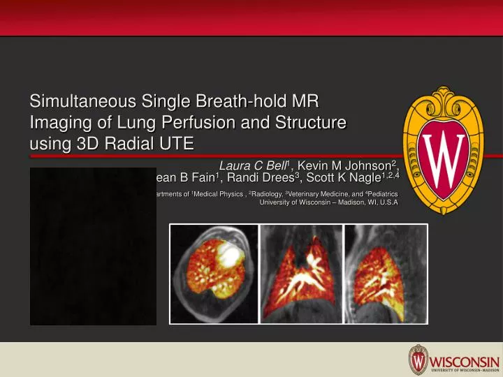
Simultaneous Single Breath-hold MR Imaging of Lung Perfusion and Structure using 3D Radial UTE. Laura C Bell 1 , Kevin M Johnson 2 , Sean B Fain 1 , Randi Drees 3 , Scott K Nagle 1,2,4. Departments of 1 Medical Physics , 2 Radiology, 3 Veterinary Medicine, and 4 Pediatrics

E N D
Simultaneous Single Breath-hold MR Imaging of Lung Perfusion and Structure using 3D Radial UTE Laura C Bell1, Kevin M Johnson2, Sean B Fain1, Randi Drees3, Scott K Nagle1,2,4 Departments of 1Medical Physics , 2Radiology, 3Veterinary Medicine, and 4Pediatrics University of Wisconsin – Madison, WI, U.S.A
Introduction • Background • Challenges in Lung Imaging • Current Structure and Perfusion Techniques • Methods • Dog Experiments • MR Protocol and Post-Processing • Results/Discussion • Future Work Outline
Many lung diseases cause structural changes in the lungs and also impair lung function such as pulmonary perfusion or ventilation. For example, • Often, structural imaging is desired to rule-out alternative diagnoses that may not manifest perfusion abnormalities • Recently, ultra short echo times (UTE) sequences have allowed for high resolution structural visualization of the lung Introduction
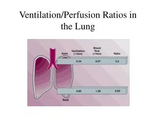
Background: Lung Imaging Challenges MR imaging of the lungs is difficult due to: 1) Cardiac and Respiratory Motion 2) Low Proton Density (0.15 g/ml) Rapid dephasing of inherently low signal. 3) Lung alveoli create local field inhomogenities and susceptibility gradients very short T2* (~1ms) †Fawcett/Gehr/Science Photo Library
Background: Current Imaging Methods • Structural lung imaging has recently benefited from the development of 3D Radial UTE sequences 1-3 • Highly accelerated contrast-enhanced perfusion MR imaging has allowed for sufficiently high temporal resolution (~1 sec) with good spatial resolution (3 – 4 mm3) typically acquired with spoiled gradient echo sequences • Therefore, evaluation of both perfusion and vascular structure require separate scans which: • Increases scan time and number of breath-holds in patients who are often short of breath • Can make it difficult to correlate the perfusion and structural abnormalities due to misregistration Cystic Fibrosis patient [1] Togao O et al, MRM (2010), v64, 1491 – 1498 [2] Kuethe DO et al, MRM (2007), v57, 1058 - 1064 [3] Johnson KM et al, MRM (2012), Optimized 3D ultrashort echo time pulmonary MRI [4] Wang K et al, J Magn Reson Imaging (2013), Pulmonary Perfusion MRI using IVD sampling and HYCR
Purpose To develop and demonstrate a high resolution breath-held 3D radial UTE acquisition to simultaneously visualize lung perfusion and structure.
Methods • Nine healthy dogs were imaged twice (separated by 2 – 4 days) resulting in 18* datasets • 8 males & 1 female • Weight 10.7 7± 1.2 kg • Mean age 13 months • Dogs were ventilated and placed in the scanner in the supine position • 3T clinical scanner • 20 elements of a commercial 32-channel phased array chest coil * The first two datasets had different scan parameters, and therefore were eliminated for analysis in this study.
Methods: Imaging Protocol • Pseudo-random temporally interleaved 3D radial UTE imaging during injection of 2.3 ml of gadobenate dimeglumine followed by a 17 ml saline flush at 2 ml/sec in the cubital vein • Dynamic 3D Radial UTE scan parameters: • Optimization of the 3D Radial UTE sequence† required no hardware modifications: • Slab-selective RF excitation with limited FOV • Variable read-out gradients • Radially oversampled projections †Johnson KM et al “Optimized 3D ultrashort echo time pulmonary MRI”
Methods: Data Reconstruction Reconstruction of 3D Radial UTE data: • IIterative sensitivity encoding algorithm (iSENSE) was used • For structural images: composite reconstruction using all 8,000 projections • For time-resolved perfusion images: temporal view-sharing method • Filter has a width of 1 sec at the center of k-space and quadratically increases to 7 sec at the edge of k-space
Methods: 3D Radial UTE Reconstruction Workflow Composite image of structure Final Reconstructed Images Raw Data One dataset of 8,000 projections Iterative Sensitivity Encoding Algorithm 33 perfusion images 33 time frames each w/ 242 projs 33 time frames each w/ ~2,000 projs Adaptive k-space filter: Width of 1 sec of center of k-space and quadratically increase to 7 sec at the edge of k-space Iterative Sensitivity Encoding Algorithm
, Methods: Data Analysis • Relative Tissue Enhancement measurements • (Max signal – Baseline signal) / Baseline signal • Mean signal determined from circular ROI placed in the right lung • Temporal Waveforms • Circular ROIs centered on pulmonary artery and aorta • Right Ventricle (RV) to Aorta Transit Times • Mean signal determined from circular ROIs centered in the RV and the descending aorta • Transit time between max signal in RV and max signal in aorta • Qualitative Relative Pulmonary Blood Flow (rPBF) maps • Calculated by indicator dilution method on a pixel-by-pixel basis †Meier P et al, J Appl Physiol (1954), 6:731-744
Coronal MIPS show first pass of contrast bolus at 1 frame/sec with 0.94 mm isotropic spatial resolution Results: Tissue Enhancement and Temporal Waveforms • Relative lung enhancement: 7.7 ± 1.5 compared to baseline • RV to Aorta Transit Times: 7.4 ± 2.0 sec
Coronal MIPS show first pass of contrast bolus at 1 frame/sec with 0.94 mm isotropic spatial resolution Results: Tissue Enhancement and Temporal Waveforms
Discussion: • Same dataset = Intrinsically co-registrated • Structural images show good depiction of airway, vessels, and lung tissue • Perfusion images show normal physiologic gravitational gradient in the A/P direction • Minimal cardiac motion due to ultrashort TE and radial sampling Results: Qualitative rPBF maps co-registered to structure Top Row: rPBF maps co-registered to structure Bottom Row: Corresponding structural images
Future Work • Co-registration of lung structure and perfusion in one breath-hold in healthy dogs is feasible: • Sufficient signal is available for structural visualization and perfusion analysis • High isotropic resolution (0.94 mm) • Acquisition is robust to cardiac and respiratory motion • To authors’ knowledge this is the first example of time-resolved 3D UTE sequence in large animals • Future work: • Application of constrained reconstruction methods (e.g. compressed sensing or HYPR) • Pulse sequence development to allow for more quantitative hemodynamic analysis • Apply this method to different lung diseases
Acknowledgements UW Madison Funding Christopher Francois Sara Pladziewicz Rebecca Johnson Grzegorz Bauman This project was supported by Clinical and Translational Science Award (CTSA) program, previously through the National Center for Research Resources (NCRR) grant 1UL1RR025011, and now by the National Center for Advancing Translational Sciences (NCATS), grants 9U54TR000021. The UW School of Medicine and Public Health from the Wisconsin Partnership Program. Check out our other UTE lung abstracts at ISMRM this year: #2005 Johnson KM, “Lung Tissue Differentiation with Magnetization Transfer Prepared Multi-Echo UTE MRI” #4538 Kruger SJ, “3D Radial Oxygen Enhanced Imaging in Normal and Asthmatic Human Subjects”