Download
1 / 145
1.46k likes | 1.62k Views
17. The Urinary System: Filtration and Fluid Balance. Multimedia Asset Directory. Slide 13 Urinary System Animation Slide 24 Renal Blood Flow Animation Slide 40 Hypovolemic Shock Animation Slide 50 Blood Loss and Blood Pressure Regulation Animation

E N D
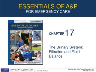
17 The Urinary System: Filtration and Fluid Balance
Multimedia Asset Directory Slide 13 Urinary System Animation Slide 24 Renal Blood Flow Animation Slide 40 Hypovolemic Shock Animation Slide 50 Blood Loss and Blood Pressure Regulation Animation Slide 51 Renin-Angiotension System Animation Slide 54 Urinalysis Video Slide 111 Renal Failure Video Slide 112 Kidney Stones Video Slide 113 Ultrasound Video
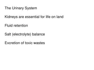
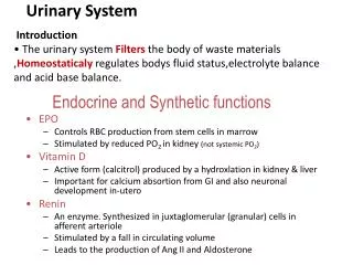
Introduction • The urinary system acts as a purification plant, cleaning the blood of waste materials. • The liver does some purification, but the urinary system controls electrolyte and fluid balances for your body. • The kidneys filter blood, reabsorb and secrete ions, and produce urine. • Without this important function you would die in a few days.
Learning Objectives • Present an overview of the organs and functions of the urinary system. • Describe the internal and external anatomy and physiology of the kidneys. • Discuss the importance of renal blood flow. • Describe the process of urine formation.
Learning Objectives • Trace the pathways of reabsorption or secretion of electrolytes and other chemicals. • List and discuss the importance of hormones for proper kidney function. • Describe the anatomy and physiology of the bladder and urine removal from the body. • Discuss several common disorders of the urinary system.
afferent arterioles (AFF er ent ahr TEE ree ohlz) aldosterone (al DOSS ter own) antidiuretic hormone (ADH) (AN tih dye yoo RET ick) atrial natriuretic peptide (AY tree al NAY tree your ET ick PEP tide) calyx, calyces (KAY licks, KAY leh seez) cortical nephron (CORE tih cull NEFF rahn) efferent arterioles (EFF er ent ahr TEE ree ohlz) Pronunciation Guide Click on the megaphone icon before each item to hear the pronunciation.
external urethral sphincter (yoo REE thral SFINK ter) glomerular capsule (gloh MAIR you ler) glomerulus (gloh MAIR yoo lus) juxtaglomedullary nephron (JUX tuh glow med DULL lair ee NEFF rahn) juxtaglomerular cells (JUX tuh glow MARE you lair) renal hilium (REE nal HIGH lum) renal medulla (REE nal meh DULL lah) Pronunciation Guide Click on the megaphone icon before each item to hear the pronunciation.
renin-angiotensin-aldosterone (RIN en-an gee oh TEN sen-al DOSS ter own) ureter (yoo REE ter) urethra (you REE thrah) Pronunciation Guide Click on the megaphone icon before each item to hear the pronunciation.
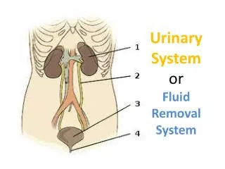
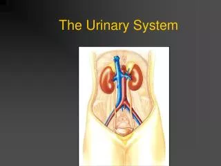
System Overview • The urinary system consists of two kidneys; bean-shaped organs located in the superior dorsal abdominal cavity that filter blood and make urine, and accessory structures. • A ureter is a tube that carries urine from each kidney to the single urinary bladder, located in the inferior ventral pelvic cavity.
System Overview • The urinary bladder is basically an expandable sac that holds urine. • The urethra is the tube that transports urine from the bladder to the outside of the body. • The job of the urinary system is to make urine, thereby controlling the body’s fluid and electrolyte balance, and eliminating waste products.
System Overview • To make urine, three processes are necessary: • Filtration – filtering the blood. What passes through the filter is called a filtrate. • Reabsorption – substances stay in the body after being removed from urine • Secretion – substances move from the blood stream and leave the body in the urine
Click here to view an animation on the topic of the Urinary System.The animation may take a moment before playing. Back to Directory
External Anatomy of the Kidney • The kidney is covered by a fibrous layer of connective tissue called the renal capsule. • The indentation that gives the kidney it’s bean-shape is called the renal hilum. • At the hilum, renal arteries bring blood to the kidneys to be filtered and renal veins take the filtered blood away from the kidney. The ureter is also attached at the hilum to transport urine from the kidney to the bladder.
Internal Anatomy of the Kidney • The kidney can be divided into three layers: • Renal cortex – outer layer – grainy in appearance and has little obvious structure to the naked eye; this is where blood filtration occurs • Renal medulla – middle layer – contains a number of triangle-shaped, striped areas called renal pyramids • Composed of collecting tubules for the urine that is formed in the kidney • Adjacent pyramids are separated by narrow renal columns – extensions of cortical tissue
Internal Anatomy of the Kidney • The kidney can be divided into three layers: • Renal pelvis – inner layer – a funnel, divided into two or three large collecting cups called major calyces. • Each major calyx is divided into several minor calyces, forming cup-shaped areas around the tips of the pyramids.
Internal Anatomy of Kidney • The blood is filtered by millions of tiny filters in the cortex, and the filtrate flows through tiny tubules in the medulla and collects in the renal pelvis. • The renal pelvis, the enlarged proximal portion of the ureter, empties into the ureter where urine is carried to the bladder.
Figure 17-2 The internal and external anatomy of the kidney.
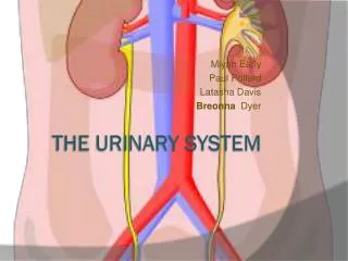
Internal Anatomy of Kidney • Blood vessels • Good blood supply to the kidney is essential to allow it to function properly – network of blood vessels throughout kidney tissue. • A single renal artery enters each kidney at the hilum, branching into five segmental arteries. • The segmental arteries branch into lobar arteries in the renal sinus.
Internal Anatomy of Kidney • Blood vessels • The lobar arteries branch into interlobar arteries which pass through the renal columns. • Arcuate arteries originate from the interlobar arteries and arch around the pyramids in the renal medulla. • Arcuate arteries give rise to cortical radiate arteries which give rise to afferent arterioles.
Internal Anatomy of Kidney • Blood vessels • Each afferent arteriole leads to a ball of capillaries called a glomerulus. • Efferent arterioles leave from the glomerulus. • Travel to a specialized series of capillaries called the peritubular capillaries and vasa recta (straight collecting tubes) that are part of the renal nephron, the functional unit of the kidney. • The peritubular capillaries wrap around the collecting tubules of the nephron, allowing efficient movement of ions between blood and the fluid in the nephron.
Internal Anatomy of Kidney • Blood vessels • From each set of peritubular capillaries, blood flows out the interlobular veins. • From there, the blood flows out a series of veins that are the direct reverse of the arteries with the exception that there are no segmental veins. • The blood finally leaves the kidney via the renal vein.
Figure 17-3 Renal blood vessels and the pathway of blood through the renal system.
Click here to view an animation on the topic of Renal Blood Flow. Back to Directory
The Nephron • The functional unit of the kidney is the nephron, consisting of millions of microscopic funnels and tubules. • The nephron can be divided into two distinct parts: • The renal corpuscle – a filter • The renal tubule – where reabsorption and secretion take place
Renal Corpuscle • Blood enters the renal corpuscle via the glomerulus, a ball of capillaries. • Surrounding the glomerulus is a double-layered membrane called the glomerular capsule, or Bowman’s capsule. • The layers of the glomerular capsule are similar to the layers of a serous membrane.
Renal Corpuscle • The inner layer of the glomerular capsule, the visceral layer, surrounds the glomerular capillaries and is made of specialized squamous epithelial cells called podocytes. This makes for a very efficient filter. • The outer layer, the parietal layer, of the glomerular capsule is simple squamous epithelium and completes the filter.
Renal Corpuscle • Blood flows into the glomerulus and everything BUT blood cells and a few large molecules, mainly protein, are pushed from the capillaries across the filter and into the glomerular capsule. • The material filtered from the blood into the glomerular capsule is called glomerular filtrate. • If blood or protein leaks into urine it can indicate a kidney filtration problem.
Renal Tubule • The rest of the nephron is a series of tubes known as renal tubules. • Glomerular filtrate travels from the glomerular capsule into the first part of the renal tubule, the proximal tubule. • The wall of the proximal tubule is made of cuboidal epithelium with microvilli.
Renal Tubule • From the proximal tubule, glomerular filtrate flows into the nephron loop (or the Loop of Henle). • The nephron loop consists of the descending loop (similar in structure to the proximal tubule) and the ascending loop (simple cuboidal epithelium).
Renal Tubule • Glomerular filtrate travels from the nephron loop to the distal tubule. • The wall of the distal tubule is like that of the ascending branch of the nephron loop. • From the distal tubule, glomerular filtrate flows into one of several collecting ducts, made of cuboidal epithelium.
Renal Tubule • The collecting ducts lead to minor calyces, then to major calyces, the renal pelvis, and the ureter. • At this point, the glomerular filtrate is urine.
Renal Tubule • Blood vessels are in close proximity to the nephrons because substances move between the tubules and the bloodstream. • Blood approaches the nephron via the afferent arteriole. • Blood flows from the afferent arteriole into the glomerulus. • Blood flows from the glomerulus via the efferent arteriole into the peritubular capillaries and vasa recta.
Renal Tubule • Blood vessels are in close proximity to the nephrons because substances move between the tubules and the bloodstream. • These surrounding blood vessels allow for reabsorption and secretion. • Blood leaves the nephron via the cortical radiate veins.
Clinical Application: Trauma, Ischemia, and Kidney Damage • The kidney is well vascularized, with each nephron surrounded by blood vessels. • The flow of blood is controlled by the afferent arteriole. When blood flow decreases for a period of time, oxygen delivery to the nephron decreases and ischemia results.
Clinical Application: Trauma, Ischemia, and Kidney Damage • Blood flow can be decreased by any number of hormonal mechanisms causing prolonged vasoconstriction, such as severe blood loss. • If the situation continues long enough, the tissues will become ischemic and eventually die, causing kidney failure that can be temporary or permanent.
From the Streets:Renal Trauma • The kidneys are well protected in the retroperitoneal space. • Trauma to the kidneys usually is the result of penetrating trauma. • Signs and symptoms
Click here to view an animation on the topic of Hypovolemic Shock. Back to Directory
Urine Formation • The kidney controls fluid and electrolyte balance by controlling urine volume and composition. • In order to form urine, the nephron must perform three processes: • Glomerular filtration • Tubular reabsorption • Tubular secretion
Filtration • During glomerular filtration, fluid and molecules pass from the glomerular capillaries into the glomerular capsule. • Across a filter composed of the walls of the capillaries and the podocytes of the glomerular capsule • The filtrate flows into the renal tubule where the chemistry is controlled by reabsorption and secretion
Filtration • During glomerular filtration, fluid and molecules pass from the glomerular capillaries into the glomerular capsule. • Filtration moves fluid and chemicals into the nephron from blood • Glomerular filtrate is chemically similar to blood
From the Streets:Kidney Stones • Kidney stones, or renal calculi, are formed by crystals in the kidney’s collection system.
Figure 17-10 Cross section through a kidney showing multi- ple stones, including one “staghorn” stone in the renal pelvis.
From the Streets:Kidney Stones • Causes • Risk factors • Signs and symptoms • Diagnostic tests • Treatment
From the Streets:Hemodialysis • Renal failure (RF) can be either acute (ARF) or chronic (CRF). • ARF is a sudden drop in urine output to less than half a liter per day. • Causes • Signs and symptoms • Treatment
Reabsorption and Secretion • Urine is chemically different from plasma because reabsorption and secretion control the concentration of chemicals and volume of urine. • Substances that are reabsorbed pass from the renal tubule into the peritubular capillaries and return to the blood stream. • Substances that are secreted pass through the peritubular capillaries into the renal tubule and eventually leave the body as urine.
Reabsorption and Secretion • Urine is chemically different from plasma because reabsorption and secretion control the concentration of chemicals and volume of urine. • Some substances, like glucose, are completely reabsorbed while substances like metabolic waste products (urea and creatinine) are almost completely secreted as urine.
Click here to view an animation on the topic of Blood Loss and Blood Pressure Regulation. Back to Directory