Download
1 / 38
770 likes | 3.43k Views
Urinary System. Functions of Urinary System: Excretion – removing nitrogenous wastes, certain salts and excess water from blood. Maintain acid-base balance Secrete waste products in the form of urine Eliminate urine from bladder. Bell. Answer questions 1-3 page 440 in text. Bell.

E N D
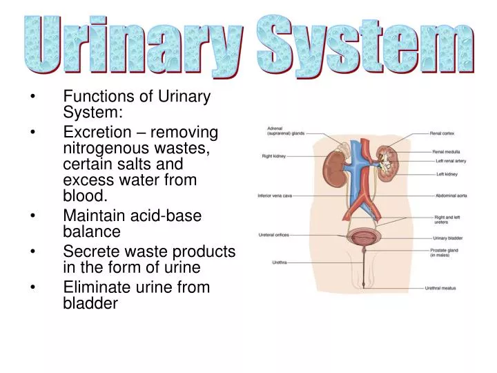
Urinary System • Functions of Urinary System: • Excretion – removing nitrogenous wastes, certain salts and excess water from blood. • Maintain acid-base balance • Secrete waste products in the form of urine • Eliminate urine from bladder
Bell • Answer questions 1-3 page 440 in text
Bell • Make sure I have all of your notes etc. from nutrition • List four main structures of the urinary system and its function
Bell… • List the 4 main structures of the urinary system
Bell • Label write on wipe off urinary system G,H, I and J only
KIDNEYS • Bean-shaped organs • Located between peritoneum and the back muscles (RETROPERITONEAL)
RENAL PELVIS • – funnel shaped structure at the beginning of the ureter (text page 402) • The renal pelvis is a reservoir for collecting urine from all parts of the kidney. Essentially a funnel, the broad end of the renal pelvis accepts the urine and channels it out of the hilus into the ureter to be discharged to the urinary bladder.
CORTEX Composed of millions of microscopic functional units called nephrons. The outer most portion of the kidney MEDULLA Inner, striated layer Striated cones are RENAL PYRAMIDS Base of each pyramid faces cortex, while apex empties into cuplike cavities called CALYCES : Two Layers
URETERS • One from each kidney • Carry urine from kidney to bladder • Smooth muscle tube with mucous membrane lining • Peristalsis pushes urine down ureters
URINARY BLADDER • Hollow, muscular organ • Made of elastic fibers and involuntary muscle • Stores urine – usually about 500cc • Emptying urine (voiding) is involuntary but controlled through nervous system (voluntary) • Urine leaves through URETHRA to outside opening = URINARY MEATUS
Build A Urinary System • Supplies you will need: • 2 Kidney beans (represents Kidney) • 2 piece of spaghetti (represents ureters) • 1 marshmallow (represents bladder) • 1 Straw piece (represents the Urethra) • 1 Blue yarn (represents the Arteries) • 1 red yarn (represents the Veins) • Making the model • 1. Glue the kidney Bean on the Diagram in the proper location • 2. The yarn will be approximately 3 inches long. Separate the yarn into 2 pieces about ½ up so t he arteries and veins can go down each leg. • 3. Insert the approximately 1 inch long spaghetti lengths into the mini-marshmallow at an angle. • 4. Insert the approximate 1 centimeter long piece of straw into the bottom of the marshmallow. This represents the urethra. Place glue on the marshmallow only. The spaghetti should be just high enough to go over the top of the yarn. • 5. Using your model describe to a partner just exactly what happens in each part of the urinary system.
Classwork • Label Structures of… • Workbook 260 C
Bell • List in order the four areas urine flows thru • What process causes the urine to pass thru tubes…wavelike?
RENAL PELVIS • – funnel shaped structure at the beginning of the ureter (text page 402) • The renal pelvis is a reservoir for collecting urine from all parts of the kidney. Essentially a funnel, the broad end of the renal pelvis accepts the urine and channels it out of the hilus into the ureter to be discharged to the urinary bladder.
CORTEX Composed of millions of microscopic functional units called nephrons. The outer most portion of the kidney. Contains proximal tubules See pg 429 MEDULLA Inner, striated layer. Contains distal tubules Striated cones are RENAL PYRAMIDS Base of each pyramid faces cortex, while apex empties into cuplike cavities called CALYCES : Two Layers
NEPHRON • Functional unit of the kidney • Most of the Nephron is located within the Cortex • Parts include: • Afferent Arteriole-Dirty Blood in • Bowman’s capsule-c shaped and surrounds the Glomerulus • Glomerulus-cluster of capillaries • Proximal convoluted tubule • Loop of Henle • Distal convoluted tubule • Collecting tubule • Efferent Arteriole-Clean blood out
Cont. labeling • Write on wipe off…all
Urine Formation in the Nephron 1- • FILTRATION • First step in urine formation • Blood from renal artery enters glomerulus • High blood pressure in glomerulus forces fluid (FILTRATE) to filter into Bowman’s capsule • Filtrate does not contain plasma proteins or RBCs – they’re too big • Bowman’s capsule filters out 125cc of fluid/min. – 7500cc/hour • As filtrate continues through nephron, 90% of water is reabsorbed • REABSORPTION • Water and useful substances are reabsorbed in the renal tubules • If blood levels of certain substances are high (glucose, amino acids, vitamins, sodium) then those substances will not be reabsorbed • SECRETION • Opposite of reabsorption • Secretion transports substances from blood into distal and collecting tubules • Substances include creatinine, hydrogen ions, potassium ions, and some drugs • Electrolytes are selectively secreted to maintain body’s acid-base balance 2- 3-
Urinary Output • Average = 1500 ml/day • URINALYSIS – examination of urine to determine presence of blood cells, bacteria, acidity level, specific gravity and physical characteristics (color, clarity and odor)
Building a Nephron • Supplies Needed • Red Yarn about 8 to 10 inches (Represents Afferent Arteriole and Glomerules) • Small Cup (Represents Bowman’s Capsule) • 1 Yellow Pipe Cleaner (twisted becomes Proximal convoluted Tubules) • Blue Pipe Cleaner (twisted becomes Distal convoluted Tubules) • 1 Hairpin (Represents Represents the Henle’s Loop) • ½ drinking straw (Represents the Collecting Tubules) • Tape and Glue • Making the Model • Using the Diagram on page 403 you are to create a nephron on a piece of paper and the materials available. While building your nephron, remember what each of the item represent. • Make sure you label the following: • 1) your nephron parts • 2) Where your nephron filtration, reabsorption, and secretion occurs.
Activity Without book or notes!Put these in order. You have 3 minutes! efferent Arteriole Distal convoluted tubule Glomerulus Proximal convoluted tubule Bowman’s capsule Afferent Arteriole Collecting tubule/duct Loop of Henle Come up to the front and check your work. If you get it right get a little treat! Put your name on your paper and leave it at the front
How did you do??? • Afferent Arteriole • Glomerulus • Bowman’s capsule • efferent Arteriole Proximal convoluted tubule • Loop of Henle • Distal convoluted tubule • Collecting tubule/duct
Bell/Payday! • Text page 440 review questions 1-8 only
Control of Urinary Secretion Chemical Control Reabsorption of H20 in distal convoluted tubule controlled by ADH (antidiuretic hormone) Secretion and regulation of ADH controlled by hypothalamus DIURETICS inhibit reabsorption of H20 Aldosterone- Secreted by the Adrenal cortex and involved in the reabsorption process Nervous Control Direct control through nerve impulses on kidney blood vessels Indirect control though stimulation of endocrine glands Renin-a hormone released when blood pressure drops
Bell • Review questions text page 440 1-8
Disorders of the Urinary System • RENAL CALCULI (Kidney Stones) • Made of crystals of calcium phosphate and uric acid • Gradually they get larger until they block ureters • First symptom – severe pain • Other symptoms – nausea and vomiting, frequency, chills, fever, hematuria • Diagnosis – by symptoms, ultrasound, or x-ray • Rx – increase fluids to flush out stone, medications, and if needed – LITHOTRIPSY
LITHOTRIPSY • Surgical procedure to remove kidney stones • Shock waves hit dense stones and break them up • Done on outpatient basis
Other urinary disorders… • NEPHRITIS • Inflammation of the kidney (kidney infection) • CYSTITIS • Inflammation of the mucous membrane lining of the urinary bladder • Most common cause – E. Coli • Symps – DYSURIA (painful urination) and frequency • Usually in females (shorter urethra) • Rx – antibiotics • INCONTINENCE – involuntary urination
DIALYSIS (HEMODIALYSIS) • DIALYSIS (HEMODIALYSIS) • Used for kidney failure • Involves the passage of blood through device with semi permeable membrane • Dialysis serves as substitute kidney • Blood from patient flows through machine and is filtered • Can be done at home or in clinic • Takes 2-4 hours, 2-3 times a week
KIDNEY TRANSPLANT • As a last resort • Involves donor organ from someone with a similar immune system • Main complication – rejection
The disorders keep trickling in….. • ENURESIS – bedwetting • GLYCOSURIA – sugar in urine • NOCTURIA – frequent urination at night • POLYURIA – large amounts of urine • PYURIA – pus in urine • ANURIA – no urine produced • HEMATURIA – blood in urine • DIURETIC – drug or substance to increase urine production
What Happened? • Read each scenario and, based on your understanding of the anatomy and physiology of the urinary system, what do you think has happened? • !) Taylor is on the track team at school. After just having run a mile on a very hot day, Taylor goes to the bathroom to urinate, and is concerned that there is a very small amount and the color is deep amber. • What happened? • 2) Carlos was playing football this afternoon and was hit pretty hard in his right flank. Tonight, he goes to the bathroom to urinate, and notices the water in the toilet bowl is a light pink. • What happened? • 3) Cara’s grandmother had a stroke and has been hospitalized. When Cara goes to visit her grandmother, there is a clear plastic bag filled with urine hanging on the side of the bed, and a tube leading from the bag to her grandmother. • What happened? • 4) Jermina goes to camp. After a few days there, she begins to have dysuria, and notices that her urine smells funny and is cloudy. • What happened?
Complete top 1/2 grid/worksheet titled Identifying Disorders of the Urinary System • Complete What Happened?
Bell… • Get this work out and place it on your desk…Complete top 1/2 grid/worksheet titled Identifying Disorders of the Urinary System • Complete What Happened? • New Assignment!...read Medical Decisions…be prepared to rank/rationale • Tear apart your nephron-save the hair pin
Bell • Make 4 strips of paper…. label A,B, C, D
Bell • I need the tests back that you studied from! • Attach the ‘Identifying Disorders of the Urinary System’ grid to your packet • Put your name on your packet and place on chair in front • Bring your write on wipe off urinary system to Lindsey