Download
1 / 54
570 likes | 757 Views
Explore the complex defense reaction of inflammation in connective tissues, involving plasma proteins, blood cells, and specific mediators. Learn about acute and chronic inflammation, inflammatory reactions, and the systemic effects on the body.

E N D
Pathophysiology of Inflammation Miklós Molnár
Essentials of Inflammation • Inflammation is a complexdefense reaction in the vascularized connective tissue in response to endogenous or exogenous stimuli in order to destroy, eliminate the injurious agents or microorganisms and initiate a series of events that heal and reconstitute the damaged tissue
Subsequent Events • Alteration (tissue injury) • Vascular reaction • Proliferation (reconstitution)
Localization • The connective tissue and its microcirculation including the surrounding extracellular space.
Participants • Plasmaproteins • Proteolytic cascades: coagulation, kinin, fibrinolytic • Blood cells • Neutrophils, eosinophils, basophils, monocytes, lymphocytes, platelets • Cells of the connective tissue • Mast cells, fibroblasts, macrophags, lymphocytes • Blood vesels • Endothelium, basement membrane, smooth muscle cells
Participants • Extracellular matrix • Fibronectin, laminin, collagen, enactin, tenascin, proteoglikansetc. • Specific mediators • Early mediatorspositive, amplification • Late mediators negative, restitutio
Injury Acute Inflammation or Restitution Chronic Inflammation or Restitution Proliferation (granuloma) Restitution
Types of Inflammatory reactions TypeAcuteChronic Inflammation Inflammation Duration max. 1 week> 1 week Characteristics exudation (edema) connective tissue emigration of leuko- proliferation cytes lymphocytes and macrophages accumulation
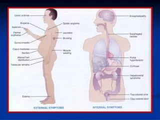
Inflammation Local reactions Systemic reactions Classical (cardinal) signs Acute phase reaction, fever, leukocytosis etc.
Celsus described the four famous signs of inflammation calor, rubor, tumor, and dolor(A.D. 30)
Systemic Effects of Inflammation Acute-phase reaction TNFa, IL-1 & IL-6 • fever • leukocytosis • Iron deficiency, anemia • proteolysis • activation of lymphocytes
g-INTERFERON IMMUNCOMPLEX NEURO- PEPTIDES MACROPHAGES ENDOTOXIN EXOTOXIN URATE SILICATE CRISTALS Initiation of Acute Phase Reaction IL-1 TNFα IL-6
Acute Phase proteins • Ceruloplasmin • Scavenges oxygen radicals generated by leukocytes • Protease inhibitors • α1-protease inhibitor, α1-antichymotrypsin and α2-macroglobulin • C-reactive protein (CRP) • binds to bacteria and produce capsular swelling, precipitation and agglutination; binding also fixes complement, thus causing the production of C3b (an opsonin) and chemotactic factors • Fibrinogen • may serve as opsonin by clumping bacteria together; breakdown products of fibrinogen has anti-inflammatory activity • Transferrin • decreases, thus limiting the amount of iron available to meet bacterial growth requirement
PATHOMECHANISM OF INFECTION, TOXINS, IMMUNCOMPLEXES, NEOPLASIA IL-1/TNF IL-6 Hypothalamus Prostaglandins (E2) Vasomotor center ? Sympathetic nerves Skin vasoconstriction ↓ Heat dissipation FEVER FEVER
Local Effect of Acute Inflammation • Blood flow increases(alteration of vascular caliber) • Increased vascular permeability(plasma proteins and leukocytes leave the circulation, retraction of endothelial cells, fenestration) • Cellular events(emigration of leukocytes and makrophages: margination, rolling, adhesion, emigration, chemotaxis, phagocytosis)
Advantages of exudation • Fluid exudation (dilution of toxins) • Increased protein content Globulins (antibodies) Fibrin precipitation (Bacterial fixation, wound healing) Acute-phase proteins
Mechanisms of Vascular Leakage 1. Endothelial contraction widening of intercellular junctions (rapid , short-lived action, histamine, bradykinin, leukotrienes etc. effects only the small venules) 2. Junctional retraction induced by IL-1, TNFα, IFN, delayed (4-6 hours) effect 3. Leukocyte-dependent leakage free radicals and proteolytic enzymes 4. Leakage from regenerating capillaries angiogenesis, intercellular junction development
Leukocyte extravasation • Margination, rolling and adhesion • Transmigration across the endothelium (diapedesis) • Migration in interstitial tissues toward a chemotactic stimulus
Adhesion receptors • Selectins • E-selectin (ELAM-1, endothelium) • P-selectin (GMP140, platelets) • L-selectin (LAM-1, leukocytes) • Immunoglobulins • endothelial adhesion molecules: intercellular (ICAM-1), vascular cell ~ (VCAM-1) both interact with integrins found on leukocytes • Integrins(transmembrane-adhesive heterodimeric glycoproteins, α and β chains) • 2-integrins (LFA-1, MAC-1 ICAM-1) • 1-integrin (VLA-4 VCAM-1)
Roles and Mechanism of Adhesion Molecules • Redistribution of adhesion molecules to the cell surface P-selectin (intracitoplasmatic granules of endothelialcells- Weibel-Palabe granules histamin, thrombin, PAFcell surfaceadhesion and rolling of leukocytes
Roles and Mechanism of Adhesion Molecules 2.Induction of adhesion molecules on endothelium (IL-1, TNF) E-selectin, and ICAM-1, VCAM-1 3.Increased avidity of binding LFA-1 on leukocytes dose not adhere to its ligand ICAM-1 on endothelium at resting condition. However, after certain stimuli LFA-1 is converted from a state of low- to high-affinity binding toward ICAM-1 Strong bindingTransmigration across endothelium
Selectins Integrins Migration of Leukocytes
Chemotaxis and Leukocyte Activation • Exogenous substances • bacterial products(N-formyl-methioninecontaining peptides, lipids) • Endogenous substances • Compounds of complement system ( C5a, C3a) • Products of the Lipoxygenase pathway (LTB4) • Cytokines (IL-8) receptor bindings PLC-Ca2+ actin-myosin active locomotion and activation of PLA2 AA, degranulation (lysosomal enzymes), modulation of adhesion molecules
Phagocytosis 1. • Recognition and attachment • opsonins mediated (Fc-fragment of IgG, C3b) (via FcR receptors) • nonopsonic phagocytosisrecognition of LPS
Phagocytosis 2. • Engulfment Binding to FcR pseudopodsfusion of the phagocytic vacules and the lysosomal granules • Killing or Degradation Oxygen dependent: NADPH oxidase superoxide ion H2O2 MPO H2O2 + Cl- HOCl NO synthase NO peroxinitrite (CONO2- ) Oxygen independent: lysozyme, lactoferrin, MBP –major basic protein-, defensins, acid hydrolase
Leukocyte adhesion deficiency 1 Leukocyte adhesion deficiency 2 Neutrophil specific granule deficiency Chronic granulomatosus disease X-linked Autosomal recessive Myeloperoxidase deficiency Chédiak-Higashi syndrome β chain of CD11/ CD18 integrins Selectin receptors (Sialyated oligosacharide) Absence of neutrophil specific granules, defective chemotaxis Decreased oxidative burst NADPH oxidase (membrane component) NADPH oxidase (cytoplasmic component) Absent MPO-H2O2 system Multiple defect Defects in Leukocyte FunctiomGenetic Disease Defect
Thermal injury, diabetes, malignancy, sepsis, immunodeficiencies Hemodialysis, diabetes Leukemia, anemia, sepsis, diabetes, neonates, malnutrition Chemotaxis Adhesion Phagocytosis and microbicidal activity Defects in Leukocyte FunctiomAcquired Disease Defect
Plasma Proteases • Kinin system • Fibrinolytic system • Complement system
The Kinin system • Bradykinin • Inducing arteriolar dilatation • Increasing the permeability of venules • 3. Causing pain • Kallinkrein • Activation of plasminogen • Activation of C1q in the complement system • Found in plasma , tissues and secretions
prourokinase urokinase Kallikrein The Fibrinolytic System Plasminogen Activating Factors • Plasmin • Produce vasodilatation by generating FDP • Can digest fibrin, removes fibrin deposit • 3. Can activate complement system by cleaving C3
Other Plasminogen Activators • Bacterial substances (eg. streptokinase) • Cell-derived activators (eg. trypsin) • Macrophage plasminogen activator • Hageman factor Note: Plasmin was formerly known as fibrinolysin, this old name is still used for this system
The Complement System • Consist of 20 interactive plasma and cell membrane proteins • Activated complement: • Mediating vascular responses (histamine release) • Requiting phagocytic leukocytes (chemotaxis) • Opsonizing target of phagocytic cells • Directly damaging target cells or tissue • Activation: • Classic pathway • Alternative pathway
Classic Pathway This pathway is activated by antibody-coated targets or antigen-antibody complexes Hageman Factor C1 inhibitor
Alternative Pathway Activated by: 1. LPS; 2. plasmin; 3. a factor from cobra venom; 4. aggregated IgM or IgG; 5. complexes of antigen with IgA C3b inhibitor
The Role of Hageman Factor C1 inhibitor
Chemical Mediators of Inflammation • Vasodilatation Prostaglandins, histamin • Vascular permeability Vasoactiv amins C3a and C5a Bradykinin Leukotrien C4, D4, E4, PAF • Chemotaxis C5a, Leukotrien B4 , lipids, bacterial products
Chemical Mediators of Inflammation • Fever IL-1; TNF, Prosztaglandin E2 • Pain Prosztaglandins, Bradykinin • Tissue injury Lysosomal enzymes (neutrophils, macrophages) oxygen metabolites
Cyclo-oxygenase Hypothesis - 1990’s membrane phospholipids INDUCTION: mitogens endotoxins cytokines (-) glucocorticoids Phospholipase A2 (-) arachidonic acid selective COX-2 inhibitors COX-1 constitutive COX-2 inducible Classical NSAIDS (-) (-) (-) Inflammatory Sites Stomach Kidney Endothelium Platelets PGE2/PGI2 PGE2/PGI2 PGI2 TXA2 PGE2 inflammatory mediators gastric cytoprotection renal blood flow haemostasis Physiological Effects Inflammation
Hierarchies of cytokines Stimulus TNF a IL-1 IL-6, IL-8, VEGF, GM-CSF