Download
1 / 53
530 likes | 684 Views
Chapter 4-Adrenal Glands. Ch. 4-- Study Guide . Critically read (1) pages pp. 61-69 before postsecretory metabolism of adrenal cortical hormones section; (2) pp. 71-76 (physiology of the mineralocorticoids) before Effects on water balance subsection.

E N D
Ch. 4-- Study Guide Critically read (1) pages pp. 61-69 before postsecretory metabolism of adrenal cortical hormones section; (2) pp. 71-76 (physiology of the mineralocorticoids) before Effects on water balance subsection. Comprehend Terminology (the text in bold/italic) Study and understand the text and corresponding figures.
§ Introduction • Adrenal hormones: • required for maintenance of life • Without them, deranged electrolyte or CHO metabolism, hypoglycemic coma, and death • Outer cortex– three steroid hormones: mineralocorticoids, glucocorticoids, and androgens • Inner medulla—a component of the sympathetic nervous system
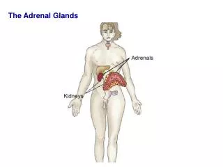
§ Morphology (1)– Fig. x + 4.1 • Location— right above the kidneys • Gross Anatomy and Histology– • Outer cortex-- > 3/4 of adrenal mass • Divided into 3 zones and produces steroids; • Zona glomerulosa– aldosterone • Zona fasciculata– cortisol and androgens • Zona reticularis– cortisol and androgens • Inner medulla -- @ 1/4 • A modified sympathetic ganglion, releases epinephrine and norepinephrine
§ Adrenal Cortex • Adrenal cortex is essential for maintenance of life. • Addison’s disease– pathological destruction or surgical removal of the adrenal cortex– death within 1-2 weeks • Why? 3 categories of hormones: Fig. 4.2– ALL come from cholesterol • Mineralocorticoids– essential to maintain sodium and potassium balance– Aldosterone + deoxycorticosterone (DOC) • Glucocorticoids– include cortisol and corticosterone– maintain CHO reserves • Androgens– on puberty and fetal life
§ adrenocortical hormones • All the adrenal steroids are from cholesterol– same as other steroids including . . . • Naming steroids— • Fully saturated 21-carbon molecule is called pregnane • Delta– location of double bond(s) and -ane changes to -ene or to –diene • Presence of a hydroxyl group (-ol) • Presence of a keto group (-one) • Fig. 4.3
All three reactions are catalyzed by a single enzyme, cytochrome P450SCC.Pregnenolone– an important molecule for other adrenal hormones
§ Adrenal Cortex (1) • Pregnenolone and progesterone (21 Carbons) is the common precursor of all steroid hormones produced by the adrenals or the gonads (Fig. 4.4)
Biosynthesis of adrenal cortical H. Z. glomerulosa & reticularis Z. reticularis; 19 carbons Z. fasciculata Z. glomerulosa
§ Adrenal Cortex (2) • A hydroxyl group at carbon 11 is found in all glucocorticoids–that is corticosterone and cortisol • Corticosterone is the major glucocorticoid in the rat but is of only secondary importance in humans • Cortisol is the most potent of the naturally occurring glucocorticoids in humans • Corticosterone is a precursor of aldosterone (a major mineralocorticoid) Fig. 4.4
Biosynthesis of adrenal cortical H. Z. glomerulosa & reticularis Z. reticularis; 19 carbons Glucocorticoids Z. fasciculata A major mineralocorticoid Z. glomerulosa
§ Adrenal Cortex (3) • Male hormones--Steroids in the 19-carbon series usually have androgenic (male hormone) activity) • Locations--This above reaction normally occurs only after puberty, and is confined to the cells of the zona reticularis (Fig. 4.4) • Female hormones--19-carbon steroids are precursors of the estrogens (female hormones; 18-carbon)—unsaturated A ring due to aromatization (loss of the methyl carbon at position 19). This reaction happens in ovary and placenta normally. Fig. 4.5
Biosynthesis of adrenal cortical H. Z. glomerulosa & reticularis Z. reticularis; 19 carbons– Male steroid hormones Glucocorticoids Z. fasciculata A major mineralocorticoid Z. glomerulosa
§ Effects of ACTH • ACTH has impact on z. fasciculata and reticularis but not glomerulosa • Through G-protein-coupled mem receptor • Increases cholesterol availability– in the cell and specifically also in mitochondria • (specifically on androgens)--ACTH is the only hormone known to control synthesis of the adrenal androgens (dehydroepiandrosterone sulfate; DHEAS) • Adrenarche– Beginning of increased secretion of adrenal hormones at puberty (another similar term: menarche) Fig. 4.6 + 4.7
§ control of aldosterone synthesis • Location– in zonaglomerulosa • ACTH is NOT an important regulator of aldosterone production in most species • Angiotensin II(from Angiotensinogen, from liver) regulates the production of aldosterone How? • (first messenger-receptor)—Angiotensin II binds with specific G-protein-coupled receptor • (second messengers)– IP3 and calcium to promote the formation of pregnenolone from cholesterol Fig. x + 4.8
§ control of aldosterone synthesis • Impact by three ions-- • (K+)-- Cells of the zonaglomerulosa are very sensitive to changes in potassium in the ECF; increased K+ (ECF) stimulates production of aldosterone • (Na+)-- Aldosterone is the principal regulator of body sodium content • (Ca+2)-- Intracellular calcium also stimulates the synthesis of aldosterone.
§ plasma binding proteins • CBG, corticosteroid binding globulin (or called transcortin), and albumin • Both are produced in the liver • CBG has a single steroid hormone binding site whose affinity for cortisol is 20 times higher than for aldosterone • About 95% of the cortisol and about 60% of the aldosterone in blood are bound to protein
4.4A. Physiology of the mineralocorticoids (Mainly aldosterone & deoxycorticosterone, also others; See Fig. 4.2)
§ Introduction • Aldosterone is the most important mineralocorticoid • Aldosterone’s physiology and life-threatening changes: • Reabsorption of sodium is decreased and fall of sodium in blood (hyponatremia) • An accompanying loss of water • Resulting decrease in blood volume called hypovolemia. • Locations of these effects– the kidney is the most important; also in the sweat glands, the colon, and the salivary glands
§ Aldosterone on the kidney-A • Increased potassium excretion • Sodium retention (decrease in urinary sodium)– The above two ions are not tightly coupled and sodium is not simply exchanged for potassium • Increase in body weight due to fluid retention Fig. 4.13
§ Aldosterone on the kidney-B • Aldosterone sensitive cells called principal cellsfound in the nephrons– specifically in the connecting tubule and the cortical portion of the collecting duct • Details— • Sodium (two-step transfer)– (A) enters the principal cells via sodium channels; (B) and is pumped out by sodium-potassium ATPase • Potassium– ROMK (renal outer medullary K+) channels on both sides of principal cells • Fig. 9.2; Fig. 4.14
In principal cells of cortical colleting duct Interstitium lumen
§ Aldosterone on the kidney-C • On principal cells– • after 30 minutes– resulting in prolonged half-life of ENaC • Later effects--Mainly by increasing the expression of proteins associated of sodium transport • Fig. 4.14B & 4.14C
In principal cells—aldosterone effects after 30 min. delay Mainly by prolonging the half-life of ENaC SGK1– serum glucocorticoid dependent kinase 1 ENaC--Epithelial sodium channel
In principal cells– later effects of aldosterone Mainly by increasing the expression of proteins associated of sodium transport.
§ Aldosterone on the kidney-D • Aldosterone also targets intercalated cells found in the nephrons– specifically in the distal nephron and collecting duct • Fig. 9.2; Fig. 4.14D
Mainly by promoting the secretion of protons (hydrogenions) in luminal membranes AR– Aldosterone receptors on the cell surface
§ Aldosterone secretion and function • Stimuli for aldosterone secretion: • Primary-- Angiotensin II • Also by ACTH and high conc. of potassium • Angiotensin II is regulated by renin from the kidney (glomerular arterioles) • Principal stimulus for renin secretion is a decrease in the blood (or vascular) volume • Principal physiology of aldosterone: • Defend the blood volume by reabsorbing sodium & water from the kidney • X + Fig. 4.15
Monitored variables– A--blood volume B--plasma potassium conc.